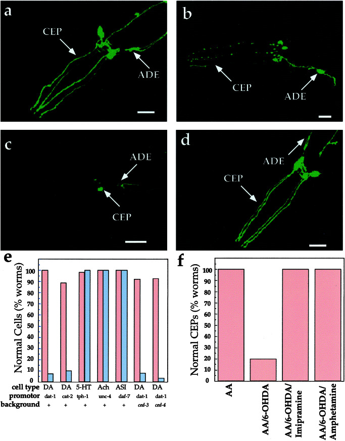
Figure 2.
Impact of 6-OHDA on dopamine and other neurons. (a) Control worms exposed to 2 mM AA. Morphological changes also were not evident with 10 mM AA. (b) Worms exposed to 50 mM 6-OHDA/10 mM AA; image chosen emphasizes the blebbing along CEP dendrites observed in animals with incomplete penetrance of the 6-OHDA effect. Similar patterns were observed at shorter time points. (c) Worms exposed to 10 mM 6-OHDA/2 mM AA. (d) Worms coexposed to 10 mM 6-OHDA/2 mM AA and 1 mM imipramine. (e) Quantitation of 6-OHDA sensitivity. Animals (≥50 worms/condition) were exposed to either 10 mM AA (red bar) or 50 mM 6-OHDA/10 mM AA (gray bar), and the GFP expression pattern in the respective cells was examined (see Materials and Methods). (f) Quantitation of DAT-1 inhibitor suppression of dopamine neuron sensitivity to 6-OHDA; animals (≥50 worms/condition) were exposed to 2 mM AA, 10 mM 6-OHDA/2 mM AA, and 10 mM 6-OHDA/2 mM AA and either 10 mM d-amphetamine or 1 mM imipramine. For all images, worms are expressing Pdat-1∷GFP. All animals were exposed to 6-OHDA as described in Materials and Methods and examined 3 days postexposure. Anterior is to the left. (All scale bars = 25 μm.) DA, dopamine; Ach, cholinergic neurons (SABV); 5-HT, serotonergic neurons (NSM); ASI, sensory neuron; +, wild type.

