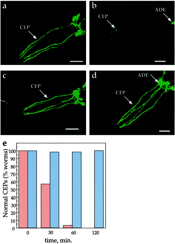
Figure 4.
Genetic suppression of 6-OHDA sensitivity of DA neurons. (a) Pdat-1∷GFP animals exposed to 10 mM AA. (b) Pdat-1∷GFP worms exposed to 50 mM 6-OHDA/10 mM AA. (c) Pdat-1∷GFP, dat-1(ok157) worms exposed to 10 mM AA. (d) Pdat-1∷GFP, dat-1(ok157) worms exposed to 50 mM 6-OHDA/10 mM AA (all animals in a–d were exposed for 2 h and examined 3 days later); (e) effect of 50 mM 6-OHDA on the CEP neurons in wild-type and dat-1 worms as a function of time. After a 2-h 6-OHDA exposure (≥50 animals per exposure condition), 100% of the worms in the wild-type background had significant degeneration in their CEPs (red bars; see Materials and Methods for scoring), whereas 100% of the dat-1 worms CEPs appear unaffected by exposure to the toxin (blue bars). (Scale bars = 25 μm.)

