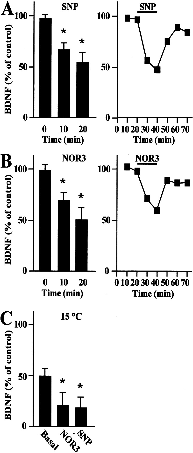
Figure 3.
Time course of BDNF secretion from primary hippocampal neurons treated with the NO donors SNP or NOR3. BDNF secretion was evaluated after the administration of 10 μM SNP (A) and 300 μM NOR3 (B) to cultured neurons. BDNF secretion was detected in the absence (0 min) or presence (10 and 20 min) of the indicated donor (Left). Values are given as mean ± SE (n = 10). * indicates statistical significance of the difference from the control at P < 0.02. In a representative experiment (Right), the respective NO donor administrated for 20 min caused a rapid down-regulation of BDNF secretion that partially recovered to baseline on withdrawal of the donor. (C) BDNF secretion evaluated after the administration of 10 μM SNP and 300 μM NOR3 at the reduced temperature of 15°C. Lowering the temperature from 37°C to 15°C caused a reduction of BDNF from basal levels that was further inhibited upon administration of SNP or NOR3 for 20 min. Values are given as mean ± SE (n = 6). * indicates statistical significance of the difference from the basal at P < 0.05.

