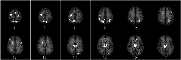
Figure 4.
Activation map overlay on female reference brain. Regions with eight or more contiguous voxels significant at level P < 0.0005 are shown. Slices are shown in radiological space (image left is brain right; image right is brain left) and are ordered from top of brain to bottom. Slice 19 is AC-PC line. See also Table 1.

