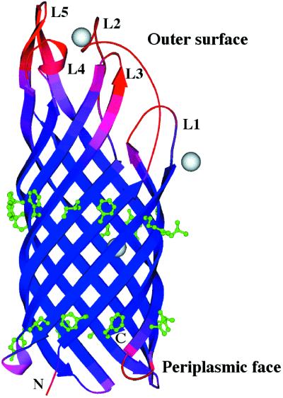
Figure 1.
Structure of N. meningitidis OpcA. The chain is color-coded for equivalent B-factors, with lower values in blue to higher values in red. Three Zn2+ ions are shown in gray, and the two rings of hydrophobic residues are shown in green. The figure was generated by using MOLSCRIPT (42).

