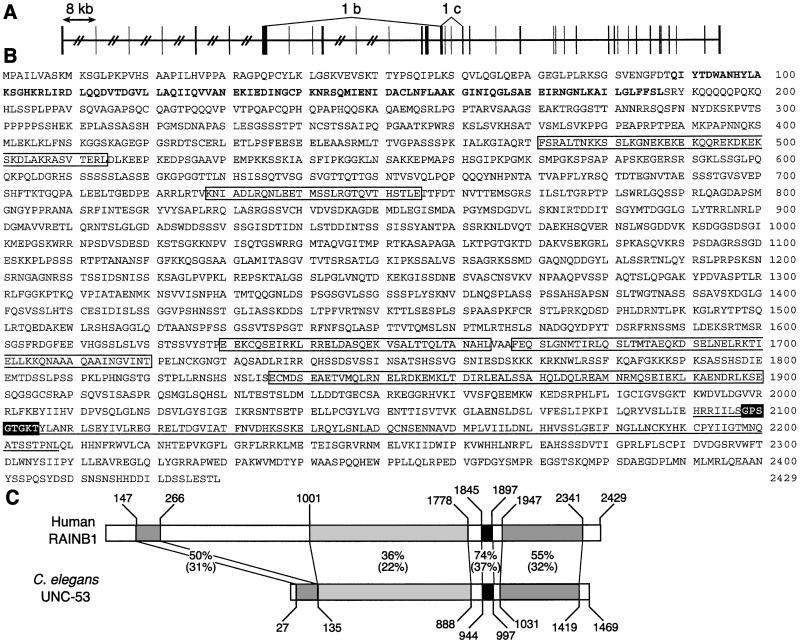
Figure 2.
Genomic organization, protein sequence, and schematic comparison of RAINB1 to C. elegans UNC-53. (A) The genomic structure of the 38 exons of the RAINB1 mRNA. The vertical boxes represent the exons, the horizontal lines represent the introns, and the hatch marks indicate introns that are greater than 8 kb. The regions encompassed by 1b and 1c are deleted in alternatively spliced transcripts identified by PCR. (B) The amino acid sequence of the RAINB1 putative ORF. The bold sequence has similarity to the calponin domain, and the boxed sequences represent putative coiled–coil domains. The white lettering with black background is a sequence motif for a P-loop ATP/GTP binding site that is contained within the underlined AAA-domain ATPase. (C) A schematic alignment of the putative RAINB1 and C. elegans unc-53 gene products. The percent similarity and identity (in parentheses) between the amino acid sequences is shown for regions indicated by shaded boxes.

