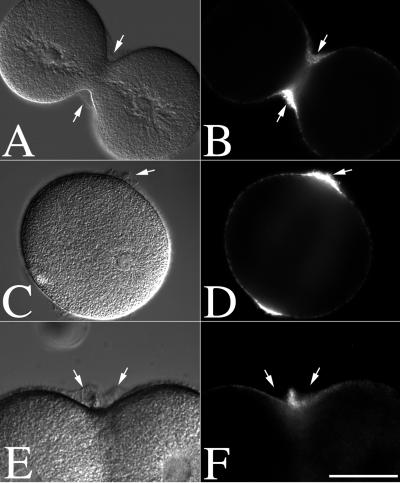
Figure 2.
Hyalin and membrane accumulation at the cleavage plane of dividing and CD-treated zygotes. L. pictus eggs were fertilized, stripped of their fertilization membranes, and cultured in CaFSW. At NEB, zygotes were transferred to normal SW (A and B) or CDSW (C–F). Cells were then fixed and processed for hyalin localization (A–D). Alternatively, cells cultured in CaFSW were transferred into CDSW containing 1 μM FM1–43 to directly visualize the plasma membrane (E and F). Arrows denote the position of hyalin. (Bar = 50 μm.)

