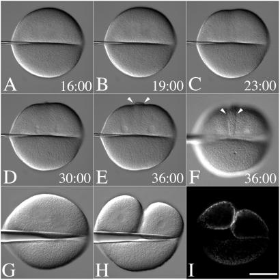
Figure 3.
Positional regulation of secretion by the mitotic apparatus. (A–F) Fertilized L. pictus eggs were cultured in CaFSW. Just before NEB, cells were transferred to CDSW. A bent microneedle was gently applied to the surface of the egg, displacing the spindle toward one side of the flattened cell. After anaphase onset (A), the astral microtubules extended toward the cortex (B), and a shallow furrow forms at the midzone between the two aster centers (C). Hyalin appears at the surface during nuclear envelope reformation (D), which accumulates further in the cleavage-arrested cell (E and F) (see Movie 1). Arrows denote the zone of membrane addition. (G–I) Just before NEB, cells were cultured on a protamine sulfate-coated coverslip. A bent microneedle was gently applied to the surface of the egg, displacing the spindle toward one side of the flattened cell (G and H). Cells were fixed in place, and after fixation, the needle was removed, and the coverslip was processed for hyalin immunolocalization (I). (Bar = 50 μm.)

