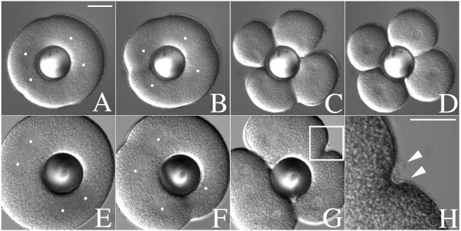
Figure 4.
Mitotic asters direct both cleavage plane determination and new membrane addition. The first cleavage of L. pictus embryos was suppressed by culturing zygotes in CaFSW containing 50 mM urethane after NEB. Shortly before NEB of the second division, cells were transferred into a chamber containing normal SW, and a glass ball was gently pressed down onto the surface of the egg, displacing the two spindles to opposite sides of the cell. In the absence of cytochalasin (A–D), cleavage furrows formed both at the midzone between overlapping asters of the same spindle, as well as between the asters of adjacent spindles. In the presence of 2 μg/ml CD (E–H), furrows initiated by overlapping asters over the metaphase plate of each spindle progressed to completion (G). Furrows induced by overlapping asters from different spindles failed to initiate or retracted (G). However, hyalin deposition at these retracted furrows was not affected (H, arrows). Dots indicate the approximate location of the aster centers. The image shown in H represents the region highlighted in G. (Bar = 25 μm.)

