Figure 5.
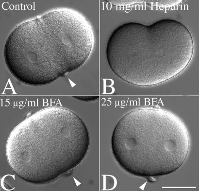
Equatorial hyalin secretion during cell division requires Ca2+-mediated exocytosis but is insensitive to BFA. (A and B) L. pictus zygotes were cultured in CaFSW until NEB, and then transferred into a chamber containing 2 μg/ml CD in normal SW. Cells were then injected with 10 mg/ml heparin, and injected cells were identified by including fluorescein-dextran in the injection buffer (not shown). Cleavage furrows were initiated in both injected and uninjected cells, but the equatorial accumulation of hyalin observed in control cells (A, arrows) was absent in heparin-injected cells (B). (C and D) To determine whether BFA affects hyalin deposition, zygotes were transferred into CaFSW containing 15 or 25 μg/ml BFA (C and D, respectively), and after NEB, transferred into CDSW containing BFA. Arrows denote the equatorial hyalin rings found at the surface of cleavage-arrested cells. (Bar = 50 μm.)

