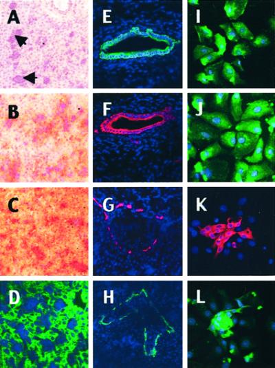
Figure 1.
Immunohistochemistry of cynomolgus monkey fetal liver for lineage-specific markers. (A–H) Liver sections (2/3 gestation). (A) Hematoxylin staining: most of the parenchyma is occupied by epithelial cells interspersed with islets of hematopoietic cells (arrows). (B–H) Immunohistochemistry with monoclonal antibodies that recognize specific simian markers as described in Materials and Methods. Two populations of epithelial cells coexist. The first type has the phenotype of hepatocytes coexpressing ALB (B, peroxidase staining and nuclear counterstaining with hematoxylin), AFP (C), and CK8/18 (D, FITC labeling and nuclear counterstaining with 4′,6-diamidino-2-phenylindole). The other type has a phenotype of biliary cells and/or progenitor cells that coexpress CK7 (E and G, Cy3 labeling and nuclear counterstaining with 4′,6-diamidino-2-phenylindole) and CK19 (F and H) in large biliary ducts and at the margin of the primitive portal tracts. Magnification, ×100. (I–L) Immunocytochemistry of freshly dissociated PFL cells (2/3 gestation). After cell dissociation, most cultured liver cells exhibit a phenotype of hepatocytes expressing ALB (I), AFP (J); clusters of proliferating cells exhibit a phenotype of biliary cells and/or progenitors cells expressing CK7 (K) and CK19 (L). Control cells and tissues did not stain specifically (data not shown). Magnification, ×400.

