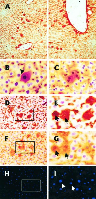
Figure 4.
Functionality of IPFLS cells after xenotransplantation in adult athymic mouse liver. IPFLS cells (1 × 10⁶) were infused via the portal vein. We visualized hepatic engraftment and differentiation by immunohistochemistry of serial frozen liver sections with monoclonal mouse antibodies that recognize simian but not mouse ALB, AFP, or CK19. (A) Liver sections were performed 8 days after transplantation and stained for simian ALB. Transplanted IPFLS cells were scattered throughout periportal areas and in the vicinity of the portal tracts. Magnification, ×100. (B–I) Liver sections were performed 21 days after transplantation. (B–G) Peroxidase staining for ALB and nuclear counterstaining with hematoxylin. (B and C) Transplanted cells have entered the hepatic parenchyma (arrow), and some of them exhibit two nuclei, a feature characteristic of adult hepatocytes. Magnification, ×400. (D–I) Immunostaining performed on serial liver section shows the differentiation pattern of the same transplanted cells (arrows). (D and E) Section immunostained for simian AFP. (F–I) Section immunostained for simian ALB (F and G, peroxidase staining) and for simian CK19 (H and I, Cy3 labeling). Magnification, ×200 and ×400.

