Abstract
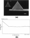
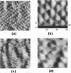
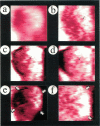
The polypeptide shell of the ferritin molecule has been imaged in water by atomic force microscopy (AFM). The central dip and the quaternary structure could be observed on the surface of the ferritin molecule anchored inhomogeneously in two dimensions. These structures observed in the AFM images are quite similar to the electron density map near the top of the apoferritin viewed down from a 4-fold axis structure reported previously (S. H. Banyard, D. K. Stammers, and P. M. Harrison, 1978. Nature (Lond.). 271:282-284). It has been achieved by introducing a "self-screening effect" of the surface charges of the AFM sample (S. Ohnishi, M. Hara, T. Furuno, and H. Sasabe. 1992. Biophys. J. 63:1425-1431) and the specially sharpened stylus of AFM cantilever.
Full text
PDF
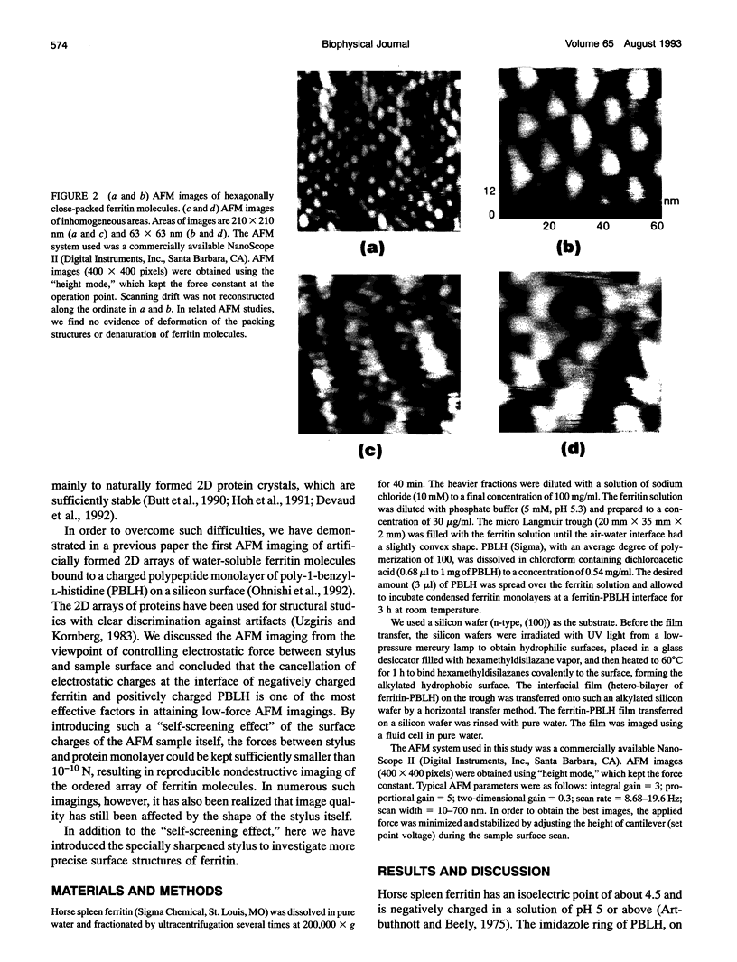
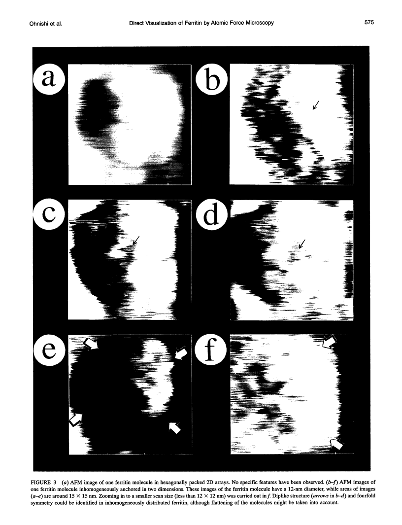
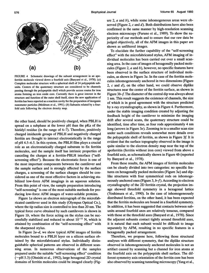
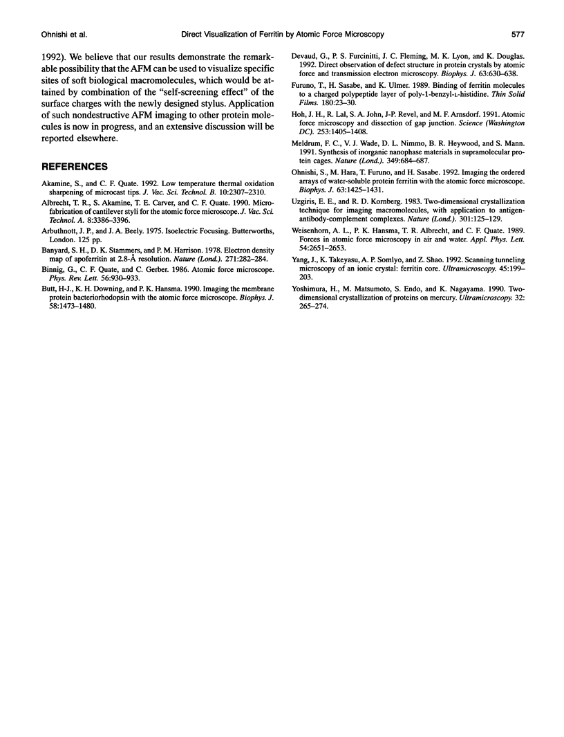
Images in this article
Selected References
These references are in PubMed. This may not be the complete list of references from this article.
- Banyard S. H., Stammers D. K., Harrison P. M. Electron density map of apoferritin at 2.8-A resolution. Nature. 1978 Jan 19;271(5642):282–284. doi: 10.1038/271282a0. [DOI] [PubMed] [Google Scholar]
- Binnig G, Quate CF, Gerber C. Atomic force microscope. Phys Rev Lett. 1986 Mar 3;56(9):930–933. doi: 10.1103/PhysRevLett.56.930. [DOI] [PubMed] [Google Scholar]
- Butt H. J., Downing K. H., Hansma P. K. Imaging the membrane protein bacteriorhodopsin with the atomic force microscope. Biophys J. 1990 Dec;58(6):1473–1480. doi: 10.1016/S0006-3495(90)82492-9. [DOI] [PMC free article] [PubMed] [Google Scholar]
- Devaud G., Furcinitti P. S., Fleming J. C., Lyon M. K., Douglas K. Direct observation of defect structure in protein crystals by atomic force and transmission electron microscopy. Biophys J. 1992 Sep;63(3):630–638. doi: 10.1016/S0006-3495(92)81651-X. [DOI] [PMC free article] [PubMed] [Google Scholar]
- Hoh J. H., Lal R., John S. A., Revel J. P., Arnsdorf M. F. Atomic force microscopy and dissection of gap junctions. Science. 1991 Sep 20;253(5026):1405–1408. doi: 10.1126/science.1910206. [DOI] [PubMed] [Google Scholar]
- Ohnishi S., Hara M., Furuno T., Sasabe H. Imaging the ordered arrays of water-soluble protein ferritin with the atomic force microscope. Biophys J. 1992 Nov;63(5):1425–1431. doi: 10.1016/S0006-3495(92)81719-8. [DOI] [PMC free article] [PubMed] [Google Scholar]
- Uzgiris E. E., Kornberg R. D. Two-dimensional crystallization technique for imaging macromolecules, with application to antigen--antibody--complement complexes. Nature. 1983 Jan 13;301(5896):125–129. doi: 10.1038/301125a0. [DOI] [PubMed] [Google Scholar]
- Yang J., Takeyasu K., Somlyo A. P., Shao Z. Scanning tunneling microscopy of an ionic crystal: ferritin core. Ultramicroscopy. 1992 Sep;45(2):199–203. doi: 10.1016/0304-3991(92)90509-i. [DOI] [PubMed] [Google Scholar]


