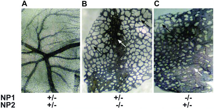
Figure 3.
PECAM staining of E10 yolk sacs shown in Fig. 2. Blood vessel yolk sacs were stained with the EC specific marker PECAM. The yolk sacs in A–C correspond to the yolk sacs in Fig. 2 D–F, respectively. (A) The NP1+/−NP2+/− yolk sac has large branching vessels and a dense capillary network. (B) The NP1+/−NP2−/− yolk sac lacks large branching vessels and lacks a capillary network. Some blood vessels are fused (white arrow). Large avascular spaces are found between the blood vessels (black arrows). (C) NP1−/−NP2+/− yolk sac. Blood vessel impairment is severe with lack of large branching vessels and capillaries, and with fused vessels (white arrow) and avascular regions (black arrow).

