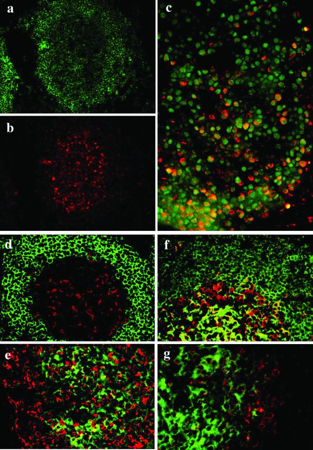
Figure 5.
Preferential expression of FREB in germinal center centroblasts. Stainings of a secondary follicle with anti-CD20 (a, green) and anti-FREB (b, red) show preferential location of FREB in large cells of the germinal center corresponding to centroblasts. (c) FREB (red) shows a perinuclear pattern of expression around the nuclear PCNA (green). Coexpression of PCNA and FREB is particularly evident at the base of the germinal center (bottom), where centroblasts are predominant. In the upper part of the germinal center, where centrocytes predominate, PCNA-positive cells are mostly FREB negative. (d–g) Expressions of FREB (red) and IgD (d), IgA (e), IgM (f), or IgG (g; green) are mutually exclusive. In f, only rare cells coexpress FREB and IgM.

