Fig 3.

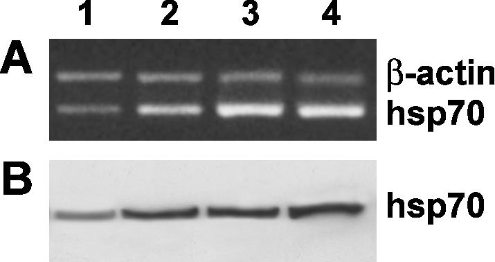
Effect of ectoine treatment on heat shock protein (Hsp)70 gene expression in keratinocyte cells. (A) Reverse transcriptase– polymerase chain reaction analysis using specific primers for hsp70 messenger RNA (mRNA) expression. Lane 1: untreated-cell mRNA; lane 2: mRNA from keratinocytes pretreated for 24 hours with 100 μg/mL ectoine, not heat stressed; lane 3: mRNA from heat-stressed keratinocytes; lane 4: mRNA from keratinocytes pretreated for 24 hours with 100 μg/mL ectoine and, successively, heat stressed. The hsp70:β-actin fluorescence intensity ratios were as follows: lanes 1, 2, 3, and 4, 0.72 ± 0.12, 1.22 ± 0.18, 2.1 ± 0.11, and 2.39 ± 0.21, respectively. The values are the mean of at least 5 independent experiments ± SD (P < 0.05). (B) Western blot analysis for the hsp70 content. Fifty micrograms of cell lysates from each sample was separated by a 12% sodium dodecyl sulfate–polyacrylamide gel electrophoresis under reducing conditions and subjected to Western blot analysis using 1 μg/mL anti-hsp70 monoclonal antibody as a probe
