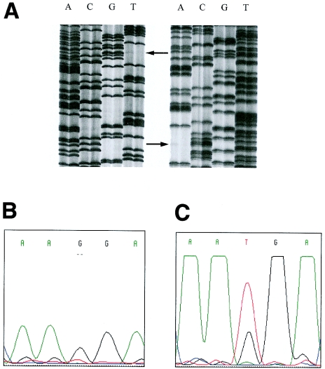
Figure 4.
A, Autoradiograph of the DNA sequence of the mosaic individual of family LA3. DNA sequences of both strands are shown (left panel shows forward sequences; right panel shows reverse sequences). Arrows indicate the mosaic mutation in the mother of the patient (left lane) in comparison with a wild-type control (right lane). B, the sequence traces of the mutated allele before mutation enrichment. C, the sequence traces of the mutated allele after mutation enrichment. Although the mutated nucleotide is not visible in B, it could be clearly identified in C, thus demonstrating that (1) standard automated sequencing analysis is not able to reveal the presence of a mosaicism and (2) that it is less sensitive than the classic radioactive sequencing protocol.

