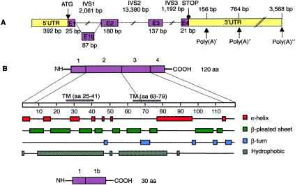
Figure 4.
Predicted schematic structure and regions of secondary structure in the human USH3 gene and in USH3 protein. A, The five exons of USH3, depicted by numbered boxes (E1–E4). The lengths of the exons are shown below the boxes, and those of the three separating introns are shown above the lines. The translation-initiation site and the first stop codon are indicated. Three polyadenylation signals (Poly(A)) and their predicted locations downstream of the termination codon are indicated by arrows. B, One-hundred-twenty-amino-acid protein, encoded by USH3, with two predicted transmembrane domains, at residues 25–41 and 63–79. The combination of putative secondary structures, such as α-helices, β-pleated sheets, and β-turns, features possible functional protein domains. An alternatively spliced transcript (bottom) of USH3 predicts a 30-amino-acid protein.

