
Figure 2. Facial photographs and neuroimaging of patients.
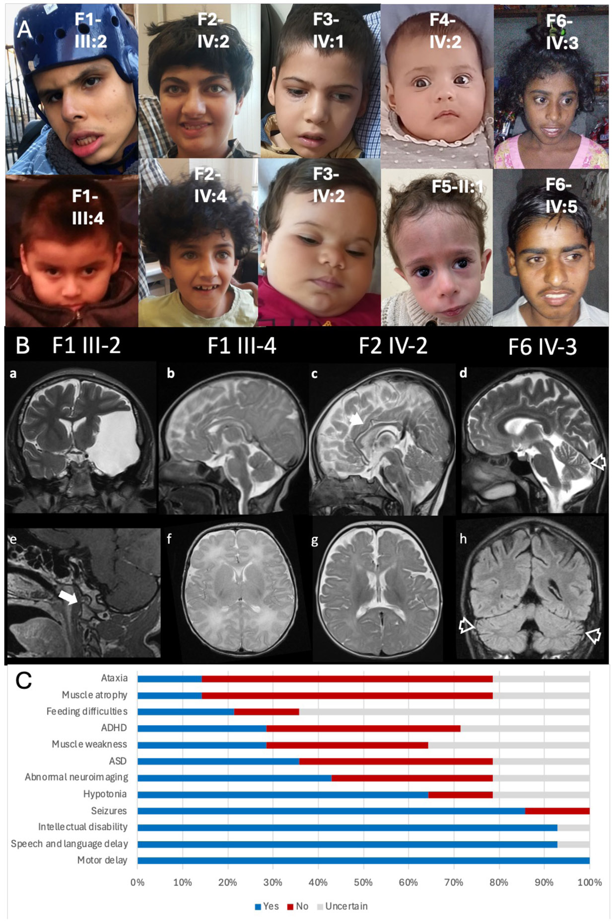
A) Facial photographs of the reported cases. B) Brain MRI. Family 1 III- 2 Coronal T2 weighted image (WI) and Sagittal post gad T1WI (a and e) shows arachnoid cyst in the left middle cranial fossa with mass effect (a) and atlantooccipital subluxation (arrow, e). Family 1 III-4 Sagittal and axial T2WI (b and e) showing normal morphology and signal changes of the brain. Family 2 IV-2 Sagittal and axial T2WI (c and g) showing mild diffuse volume loss with slightly thin corpus callosum (arrowhead, c). Family 6 IV-3 Sagittal T2WI and coronal FLAIR (d and h) showing brain volume loss including involvement of the cerebellar vermis and cerebellar hemispheres (open arrowheads, d and h). C) Common clinical features exhibited by patients, demonstrated by percentage affected.