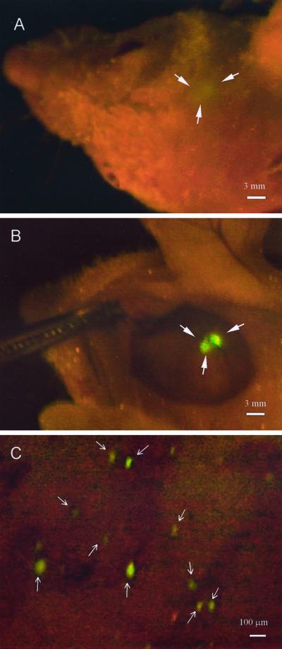
Figure 1.
Whole-body and direct view of macro and microfocal tumors in the brain. (A) Whole-body external imaging through the scalp of the bolus of U87-GFP tumor cells immediately after inoculation of tumor cells. The imaged area was ≈2 mm in diameter. (Bar = 3 mm.) (B) The scalp-flap window enabled direct view of the bolus of U87-GFP tumor cells immediately after inoculation of tumor cells. The imaged area was ≈2 mm in diameter. (Bar = 3 mm.) (C) Direct view of single tumor cells that separated from the bolus via the scalp-flap window. (Bar = 100 μm.) See Materials and Methods for details.

