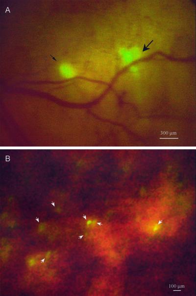
Figure 3.
Direct view of colon cancer liver micrometastasis. (A) Ventral direct view of micrometastasis of AC3488-GFP in the right lobe of the liver via a skin-flap window over the upper abdominal wall at day 7 after SOI. The smaller micrometastasis is ≈150 μm in diameter (fine arrow), and the larger is ≈300 μm in diameter (thick arrow). (B) Direct view of single cells (arrows) in right lobe of the liver after intraportal injection of 106 Colo-320 GFP cells. (Bar = 100 μm.) See Materials and Methods for details.

