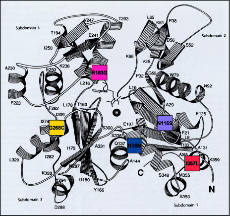
Figure 1.
Position of mutated residues in α-skeletal actin. Five different missense mutations were identified in seven patients with NM. These mutations' respective locations within the actin molecule are represented by a schematic model of the three-dimensional structure of actin (Kabsch et al. 1990, p. 37 [reprinted by permission from Nature]). Patient 1 (I357L) (orange) shows lethal severe congenital; patient 2 (R183G) (pink), lethal severe congenital; patient 3 (G268C) (yellow), childhood onset; patient 4 (I136M) (blue), typical congenital. Also represented is the autosomal dominant family (N115S) (purple).

