Figure 5.
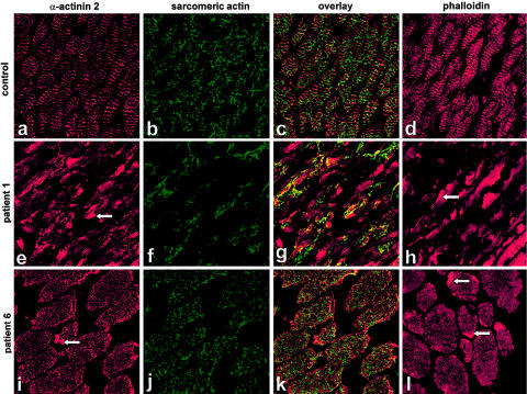
α-Actinin 2, sarcomeric-actin, and phalloidin staining of muscle-biopsy sections from patients 1 and 6. Immunolabeling with α-actinin 2 both demonstrated intense positive staining of Z-lines in all the patients and intensely stained the rods (arrowheads in panels e and i). Muscle sections labeled with a sarcomeric-actin antibody reveal a meshlike honeycomb staining pattern in the control (b) and in mildly affected patients (as shown for patient 6, in panel j). The two severely affected patients showed abnormal localization of actin, with large areas devoid of staining (as shown for patient 1, in panel f). Double labeling with α-actinin 2 and α-skeletal actin (g) demonstrated that areas devoid of sarcomeric actin in patients 1 and 2 contained α-actinin 2–reactive striations. Unlike the sarcomeric-actin antibody, phalloidin demonstrated a striated staining pattern and intensely stained the rods (arrowheads in panels h and l).

