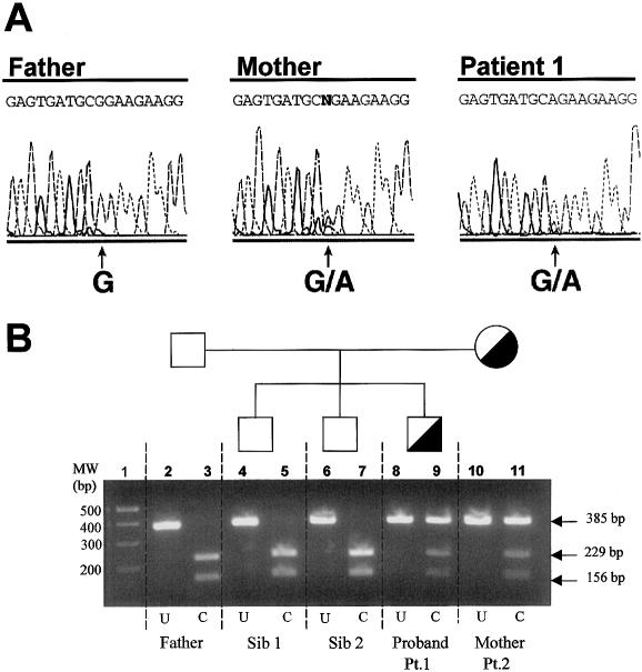
Figure 4.
Molecular diagnosis of sialuria in the family of patient 1. A, Genomic DNA sequencing of patient 1, his father, and his mother (patient 2). At position 848, the father has the normal G, but both the patient and his mother are heterozygous for the G→A substitution. B, Agarose gel electrophoresis of reaction products formed by AciI restriction-enzyme digestion of a 385-bp fragment that includes nucleotide 848. Lane 1, molecular weight markers. Lanes 2 and 3, father’s DNA, uncut (U) and completely cut (C) into 229-bp and 156-bp fragments. Lanes 4–7, DNA from the two unaffected siblings, uncut (lanes 4 and 6) and completely cut (lanes 5 and 7). Lanes 8–11, DNA from the proband (patient 1) and his mother (patient 2), uncut (lanes 8 and 10) and cut (lanes 9 and 11). The cut DNA shows heterozygosity for the normal sequence (229-bp and 156-bp fragments) and the mutation (uncut 385-bp fragment).

