Figure 1.
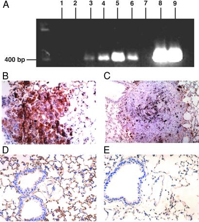
(A) RT-PCR analysis of anti-NF-κB ribozyme expression in tumor-bearing mice. Lanes 1 and 2, metastatic lung tumors from untreated mice; lanes 3 and 4, tumors from CLDC-p65-R-treated mice; lanes 5 and 6, tumors from CLDC-p50-R-treated mice; lane 7, distilled water; lanes 8 and 9, p65-R and p50-R plasmid DNA, respectively. (B) Photomicrograph (40× ocular) of p65 immunostaining of lung from the pVector-treated control demonstrates pulmonary parenchyma with a centrally placed metastatic melanoma tumor containing black, granular melanin pigment within the cytoplasm. Tumor shows golden-brown immunopositive staining for p65. (C) Photomicrograph (20× ocular) of p65 immunostaining of lung from the p65-R-treated group demonstrates a metastatic melanoma tumor in which immunostaining for p65 is essentially negative. This tumor also demonstrates some black melanin pigment similar to A. (D) Photomicrograph (40× ocular) of p65 immunostaining of lung from pVector-treated (control) group. Respiratory epithelium shows staining of some cells. Vascular endothelium is strongly positive. Alveolar epithelium also demonstrates moderate to intense reactivity of the majority of cells. Vascular endothelial cells and alveolar epithelial cells were identified by using classic microscopic criteria. We have previously shown that endothelial cells are positive for immunoreactivity using antifactor VIII antibodies and negative using anti-cytokeratin antibodies AE1/AE3, with a reverse expression pattern for alveolar lining cells (14). (E) Photomicrograph (40× ocular) of p65 immunostaining of lung from the p65-R-treated group. Respiratory (pseudostratified, ciliated) epithelium, stromal cells, and vascular endothelium are largely negative for p65 immunostaining. Alveolar lining cells show minimal, focal reactivity of low intensity (golden-brown).

