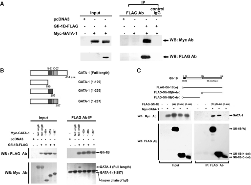
Figure 6.
Gfi-1B interacts with GATA-1. (A) Expression vector of Myc-GATA-1 together with either the control (C) or Gfi-1B-FLAG expression vector was transfected into 293T cells. After transfection for 24 h, whole cell lysates were prepared and immunoprecipitated with anti-FLAG (M2) beads or control IgG. The immunoprecipitates were subjected to immunoblot analysis with anti-Myc antibody. The blot was reprobed with anti-FLAG (M2) antibody to confirm that Gfi-1B was successfully immunoprecipitated. The input indicated 10% of the whole cell lysate used for the co-immunoprecipitation. (B) Schematic representation of wild-type and deletion constructs of GATA-1 (upper panel). N-ZF indicates N-terminal zinc finger; C-ZF, C-terminal zinc-finger. Various deleted constructs of Myc-GATA-1 were co-transfected with Gfi-1B-FLAG into 293T cells. Cells were harvested for immunoprecipitation, and the immunoprecipitates were subjected to immunoblot analysis with anti-Myc (9E10) and anti-FLAG (M2) antibody as described above. (C) Schematic representation of deletion constructs of Gfi-1B (upper panel). Control vector, wild type, N-del (164–330) or C-del (1–137) deletion construct of FLAG-Gfi-1B was costransfected with Myc-GATA-1 into 293T cells. Cells were harvested for immunoprecipitation and the immunoprecipitates were subjected to immunoblot analysis with anti-Myc (9E10) and anti-FLAG (M2) antibody.

