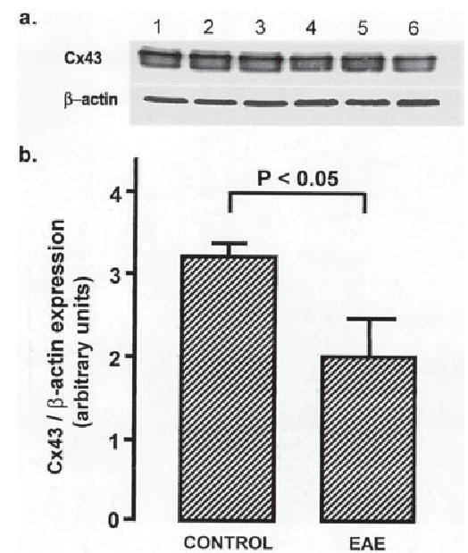
Fig. 4.

Cx43 protein is reduced in spinal cords of mice with EAE. a: A Western blot demonstrating reduced Cx43 protein in lumbar spinal cord homogenates from mice with EAE (lanes 4–6) compared with controls (lanes 1–3). b: A graph showing semiquantitative analysis based on Cx43 to β-actin ratios (mean ± SEM). There is a significant reduction of Cx43 in EAE (control 3.17 ± 0.12; EAE 1.99 + 0.41; n = 3; P < 0.05, ANOVA followed by Student’s t-test).
