Figure 2.
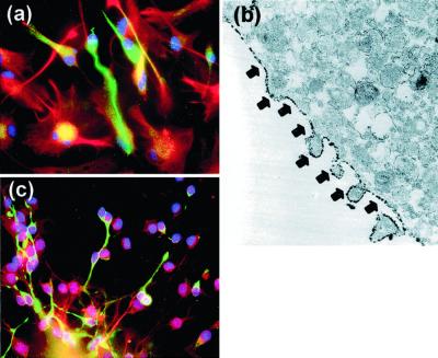
Expression of reelin and α3-integrin receptor subunits in cultures of HNSCs. (a) Reelin immunoreactivity (green) expressed by neuronal-like phenotypes and not by GFAP-positive cells (red) (×400). (b) Electron-microscopic image (×17,500) of α3−integrin receptor subunit immunoreactivity located on HNSC plasma membrane (arrows). (c) α3-Integrin receptor subunit immunoreactivity (green) expressed specifically by neuron-like phenotypes (×400). Nuclei were counterstained with DAPI (blue).

