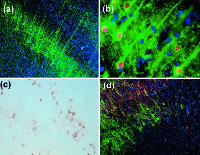
Figure 4.
Immunohistochemistry of cerebral cortex in a wild-type mouse 4 weeks after HNSC transplantation into the brain. (a) HNSCs migrated into the cortex and differentiated into neuron-like phenotypes expressing βIII-tubulin immunoreactivity (green) (×100). (b) Pyramidal neuron-like phenotypes, with the presence of apical dendrites (green) expressing BrdUrd-positive nuclei (red) (×400). (c) Note a layer of GFAP-positive HNSCs (brown) (×100). (d) Double GFAP and βIII-tubulin immunostaining revealing a layer of GFAP-positive (red) and a layer of βIII-tubulin-positive (green) cells (×100). Nuclei were counterstained with DAPI (blue).

