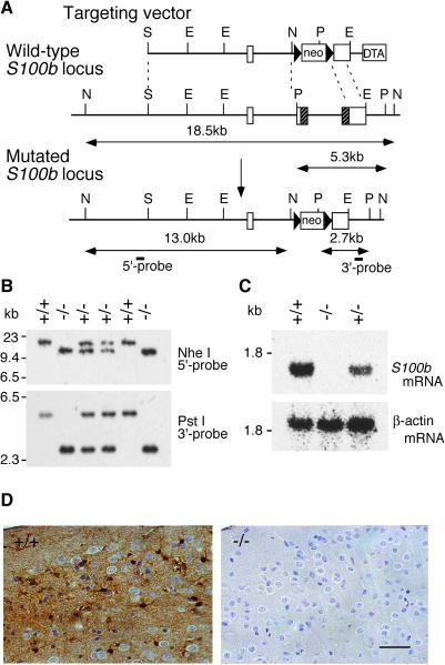
Figure 1.
Targeted disruption of the S100b gene. (A) Schematic representation of the targeting vector, wild-type S100b locus, and mutated S100b locus. Boxes in the wild-type allele denote exons 1–3. Shaded parts of exons 2 and 3 correspond to protein coding regions. E, EcoRI; N, NheI; P, PstI; S, SalI. (B) Southern blot analysis of progeny from a cross of F1 heterozygotes by using the probes indicated in A. (C) Northern blot analysis of brain total RNA (20 μg) from wild-type (+/+), heterozygous (+/−), and homozygous (−/−) mice by using a murine S100b protein coding sequence (Upper) and β-actin (Lower) as probes. Mutant mice completely lack S100b mRNA. (D) Immunohistochemistry of the cerebral cortex with an anti-S100B monoclonal antibody. The sections were lightly counterstained with hematoxylin to indicate cell nuclei. Soma and processes of cortical astrocytes were stained in wild-type mice (Left). There was no detectable immunoreactivity observed in mutant mice (Right). (Scale bar, 50 μm.)

