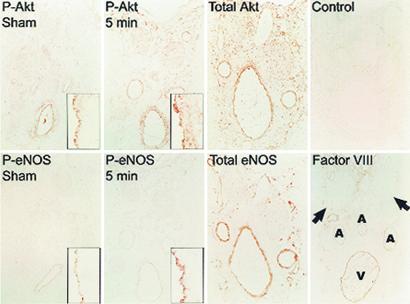
Figure 2.
Immunohistochemical localization of Akt and eNOS in serial sections of rat penis shows increased staining of phosphorylated forms in the endothelium after electrical stimulation. Rats were sham-treated or electrically stimulated for 5 min before excising the penis and staining for the indicated proteins. Low-power views (×40) show a cross-section of the penis with major structures marked in the last micrograph. (Insets) Segments of penile vascular endothelium stained for phospho-Akt or phospho-eNOS (×400) are shown. Factor VIII staining is specific for the endothelium. A, artery; V, dorsal vein. The arrows indicate dorsal nerves.

