
Figure 6.
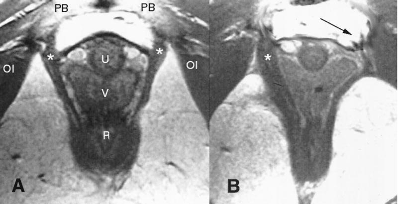
(A) Normal anatomy in an axial midurethra proton density magnetic resonance image showing the pubovisceral muscle (*) (see Figure 1 for orientation). (B) Woman who has lost a part of the left pubovisceral muscle (displayed on the right side of the image, according to standard medical imaging convention) with lateral displacement of the vagina into the area normally occupied by the muscle. The arrow points to the expected location of the missing muscle. The puborectalis is left intact bilaterally. OI = obturator internus; PB = pubic bone; U = urethra; V = vagina; R = rectum.
Lien. Muscle Stretch During Birth. Obstet Gynecol 2004.