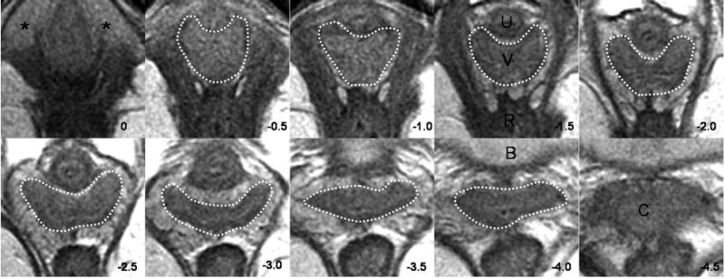
Fig. 1.

Axial slices at 5-mm intervals arranged caudal to cephalad starting from the image in the upper left (image 0). Vaginal tracings were made from above the level of the vestibular bulbs (VB), represented by asterisks (*), caudally (image 0) to below where the cervix (C) could be seen (image -4.0). U, urethra; V, vagina; R, rectum; B, bladder. © DeLancey 2004.
Hsu. Vaginal Thickness and Pelvic Prolapse. Obstet Gynecol 2005.
