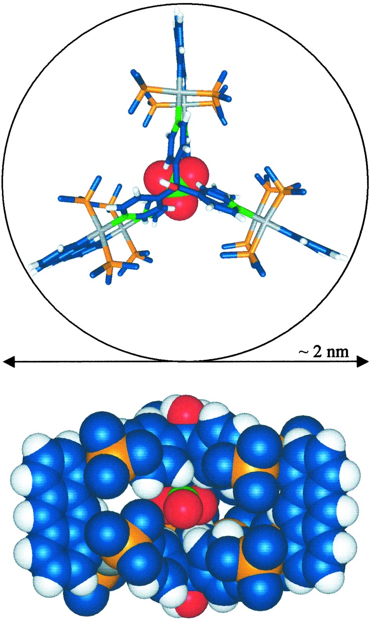
Figure 1.
Two perspectives of the x-ray structure of 3a. (Upper) The framework is shown as a stick model, and the encapsulated nitrate is represented with a space-filling model. All other counterions and the methyl groups from the triethylphosphines are omitted for clarity. (Lower) Space-filling model viewed down the C2 axis. Colors: C, blue; H, white; N, green; O, red; P, orange; Pt, gray.

