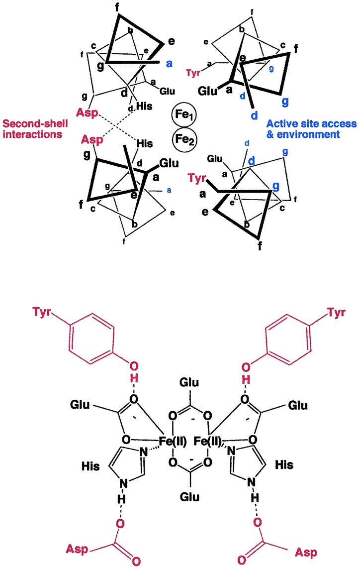
Figure 1.
(Upper) Schematic view of a 12-residue slice through the active site of the crystal structure of active site of DF1. The diagram is based on the crystallographic structures of di-Mn(II) (9) and di-Zn(II) (7) derivatives of DF1. Second-shell hydrogen bonds are shown in red, and some of the positions that help define the environment of the active site are in blue. (Lower) Schematic of the first- and second-shell ligands defining the metal-binding site (this diagram is meant to describe the atomic connectivities but not the precise geometry of the site).

