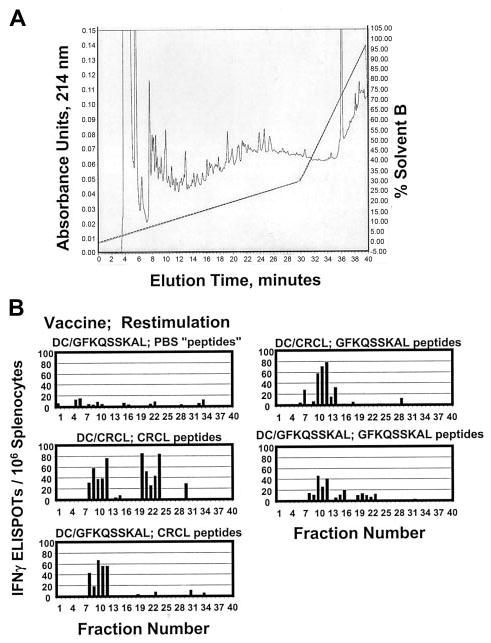
Figure 2.

Fractionation of antigenic peptides in 12B1 CRCL. (A) Peptides were acid-stripped from 12B1 CRCL and applied to a reverse-phase C18 HPLC column (rpHPLC). Peptides were separated with a 0% to 30% acentonitrile gradient over 30 minutes, followed by a 30% to 100% acetonitrile gradient over the next 10 minutes (40 minutes total, dotted line shows solvent B gradient). Elution was monitored at 214 nm (shown from 0–0.15 arbitrary units). In addition, GFKQSSKAL peptide was likewise subjected to HPLC (data not shown). Phosphate-buffered saline (PBS) was treated identically to CRCL as a faux peptide separation and as a negative control (data not shown). (B) Forty HPLC fractions from each peptide separation were collected, dried, washed, reconstituted in media, and used to restimulate the splenocytes from mice vaccinated with DC/GFKQSSKAL or DC/12B1 CRCL. IFN-γ production was determined by ELISPOT assay. PBS “peptides” refers to materials derived from PBS and fractionated by rpHPLC.
