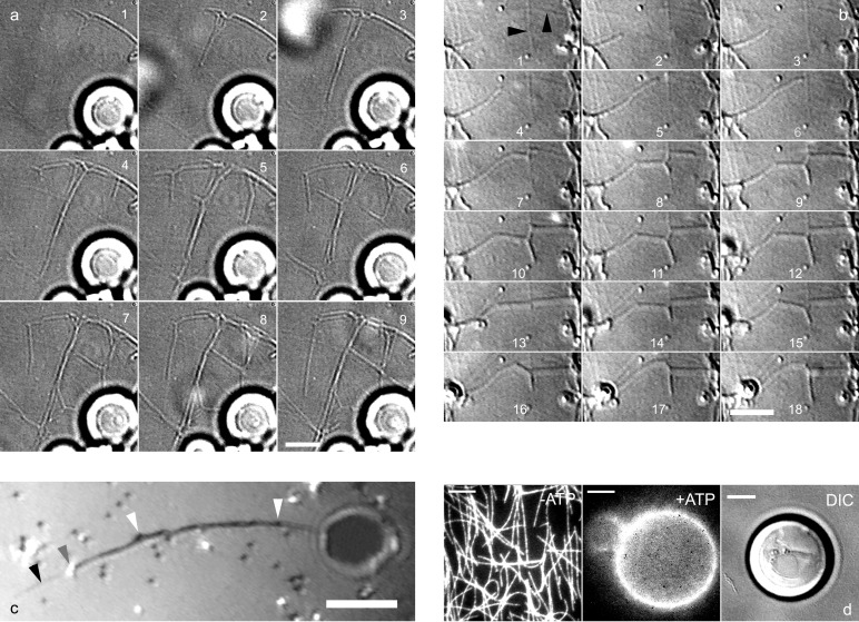
Figure 1.
Formation of tubes and networks from EPC GUVs. (a) DIC images recorded with a charge-coupled device camera (time lapse, 12 s) showing several tubes growing simultaneously from GUVs (bright spherical objects visible at the bottom of the image). Branching events occur on images 1, 4, and 5, leading to the formation of a network (images 6–9). (b) DIC images (time lapse, 7 s) showing a tube growing along a microtubule that splits into 2 tubes at the intersection of 2 microtubules (image 7). A retraction event is visible on images 12 and 13, followed by further growth (images 14–18). Black arrows point to microtubules. (c) Tube growth observed by reflection interference contrast microscopy. Beads appear black or white depending on their distance to the substrate. Two beads (gray arrow) are visible at the tip of a growing tube. Beads are also present along the tube (white arrow) growing on a microtubule (black arrow). (d) Tubes do not form in the absence of beads. From Left to Right: without ATP, biotinylated kinesins-fluorescent Cy3 streptavidin complexes were fixed on the microtubule network. In the presence of 1 mM ATP, the complexes were transferred onto the GUV (Center). Absence of tubes growing from a GUV as shown by DIC microscopy (Right). [Bar = 5 μm.]

