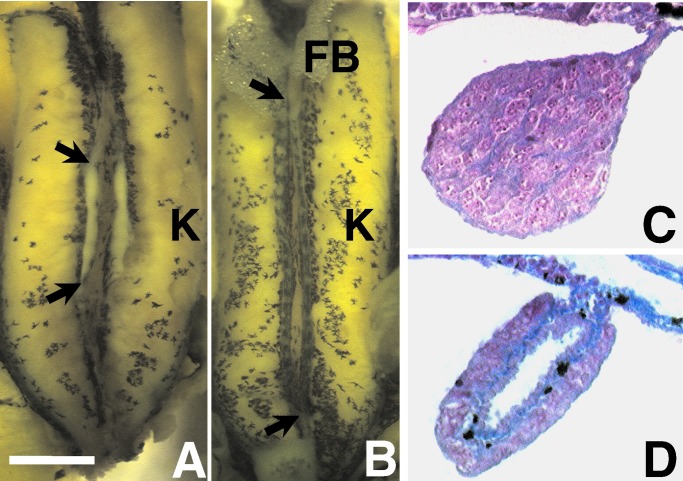
Figure 1.
Gonads of a control postmetamorphic male (A and C) and female (B and D) X. laevis. A and B show the entire dissected kidney–adrenal–gonadal complex preserved in Bouins' fixative. C and D show 8 μm of transverse cross-sections through the animals' right gonad stained with Mallory's trichrome stain. [Bar = 0.1 mm (A and B) and 10 μm (C and D)]. FB, fatbody; K, kidney. Arrows (in A and B) show the anterior and posterior ends of the animals' right gonads. The yellow color in A and B is a result of fixation in Bouins' fixative. Without fixation, the gonad is transparent. The ovary is distinguished by its greater length, lobed structure, and melanin granules. Although some specimens' ovaries lack pigment (especially atrazine-treated animals), testes never have melanin in this species. Histologically, the ovary is distinguished by the ovarian vesicle (hole in the center) along its entire length and the internal ring of connective tissue (in blue). Note the melanin granules (black) in the connective tissue in D.

