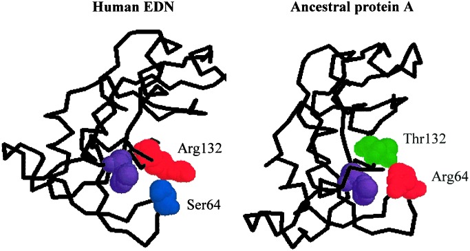
Figure 4.
Crystal structures of human EDN and ancestral protein A, showing the physical interaction between residues at positions 64 and 132. The protein backbones are shown with the spacefill presentation of the residues at the two important sites as well as that of the catalytic His-129 (in purple). The structure of the ancestral protein A was modeled according to the human EDN structure (PDB Id: 1HI2) by SWISS-MODEL (53). Both structures are depicted with the RASMOL program (54).

