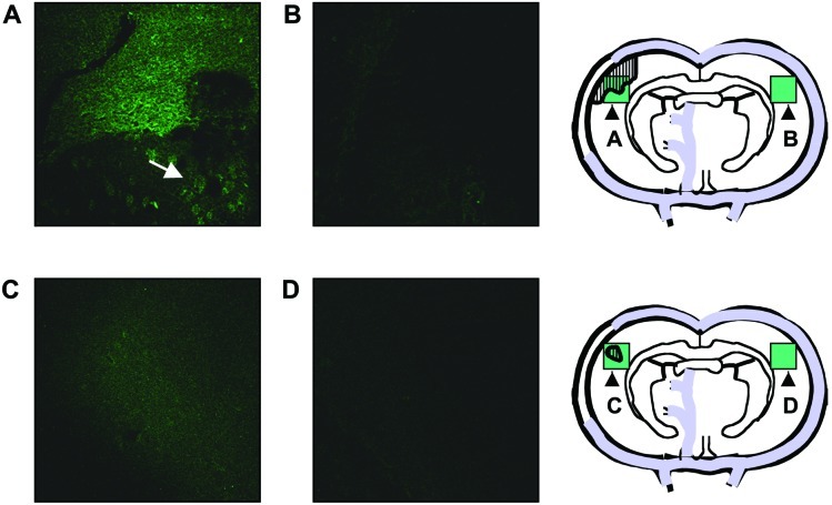
Figure 3.
N-2-MPG reduces 3-aminopropanal production during cerebral ischemia. Experimental MCAO, and N-2-MPG treatment where indicated, were performed as described in Materials and Methods. At 24 h, the brains were removed, and a section of the brain corresponding to the area of MCAO was sectioned and stained by immunofluorescence using antibodies against 3-aminopropanal-modified proteins as described in Materials and Methods. Pictures taken are representative of observations from 3–4 animals/group. All pictures were taken at ×10 magnification. (A) Detection of intense immunofluorescence for 3-aminopropanal-modified proteins in the anatomic region of cerebral ischemia in the ischemic hemisphere. The arrow shows intracellular 3-aminopropanal-modified protein fluorescence of scattered pattern in the ischemic penumbra. (B) 3-Aminopropanal-protein immunofluorescence was not detected in the opposite, nonischemic brain hemisphere. (C) N-2-MPG treatment reduces the overall area and intensity of 3-aminopropanal-modified protein immunofluorescence. (D) 3-Aminopropanal-protein immunofluorescence was not detected in the opposite, nonischemic brain hemisphere of N-2-MPG treated animals. Schematic diagrams designate in green the approximate area and location of the fluorescent pictures shown, and the hatched areas represent the approximate area of intense immunofluorescence for 3-aminopropanal-modified proteins.

