
Figure 1.
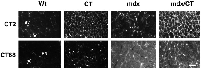
Expression of the CT carbohydrate antigen and the CT GalNAc transferase in transgenic (CT) and dystrophic (mdx) skeletal muscles. Skeletal muscles from 5-week-old mice that overexpress the CT GalNAc transferase (CT) in a wild-type (Wt) or mdx background were compared. Sections of gastrocnemius muscle were stained with antibodies to the CT antigen (CT2) or the CT GalNAc transferase (CT68) and costained α-bungarotoxin (not shown) to label neuromuscular junctions. CT2 stained small blood vessels (BV) and neuromuscular junctions (arrow) in Wt mice, but was expressed along myofibers in CT and mdx/CT transgenic mice. CT GalNAc transferase (CT68) was expressed at neuromuscular junctions and in peripheral nerves (PN) in Wt muscles. Most myofibers expressed the CT GalNAc transferase in CT and mdx/CT animals, whereas diffuse intracellular staining was also present in mdx and mdx/CT mice. (Bar = 25 μm.)