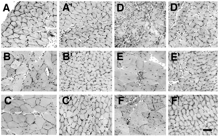
Figure 2.
Comparison of skeletal muscles from mdx and mdx/CT animals. Sections of diaphragm (A and D), quadriceps femoris (B and E), and tibialis anterior (C and F) from mdx (A–F) and mdx/CT (A′–F′) animals were stained with hematoxylin/eosin at 5 weeks (A–C) or 6 months (D–F) of age. All muscles taken from mdx animals, with the exception of 5-week-old diaphragm, displayed increased central nuclei and variability in myofiber diameter. In addition, areas of necrosis and some regions of fibrosis were seen in 6-month-old mdx muscles, especially in the diaphragm. By contrast, muscles from mdx/CT animals displayed none of these characteristics of dystrophic muscles. (Bar = 25 μm.)

