
Figure 5.
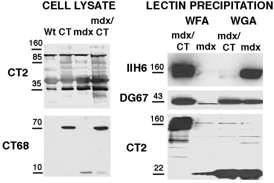
Characterization of CT and CT GalNAc transferase expression and glycosylation on α-dystroglycan with CT in mdx/CT muscles. 20 μg of protein from whole-cell lysates from wild-type (Wt), transgenic (CT), mdx, and mdx/CT muscles (gastrocnemius) were blotted with antibodies to the CT antigen (CT2) or the CT GalNAc transferase (CT68). For lectin precipitation, 150 μg of protein from nonionic detergent extracts of mdx and mdx/CT muscles was precipitated with WFA- or WGA-agarose. WFA binds to βGalNAc residues such as those on the CT antigen, while WGA typically binds GlcNAc. Precipitated proteins were then blotted with antibodies to α dystroglycan (IIH6), β dystroglycan (DG67), or the CT antigen (CT2). In cropped lanes, no other bands were present.