
Figure 3.
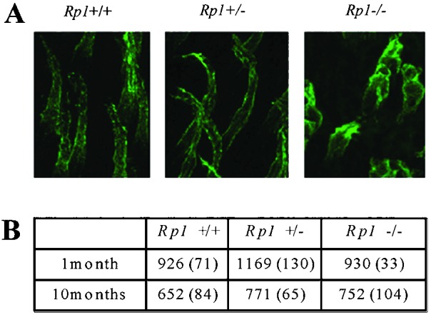
(A) Confocal microscopic images of cone OS and inner segment in Rp1 mutant retinas at 10 months of age. Whole-mount retinas were stained with peanut agglutinin, which binds specifically to cone OSs and inner segments. Serial transverse photomicrographs of retinas from each genotype were taken and superimposed. (B) The number of cones in the retinas of the three genotypes of Rp1 mutant mice at 1 and 10 months of age. From each retina, four different 250 × 250-μm areas (one from the periphery, one from the center, and two between the center and periphery) were selected, and the number of stained cones was counted. The mean values and SDs (in parentheses) are shown.