



Abstract
Since its introduction by Cravatt and colleagues in 1999, activity-based protein profiling (ABPP) has become widely utilized throughout academia, government, and industry laboratories to study enzymes spanning numerous gene families in a multitude of biological systems. As a variation of ABPP, competitive ABPP provides a powerful approach to characterize the binding behavior of small molecule probes and clinical drugs throughout the functional proteome. The power and flexibility of competitive ABPP are exemplified by a wide range of creative adaptions which increase assay throughput, enable diverse detection schemes, and support the implementation of this approach within a hybrid target-based screening platform. We review major developments in competitive ABPP through a compare-contrast format to provide a useful introduction to this enabling technology for scientists in chemical biology and drug discovery.
1. Introduction
A major challenge in modern biomedical research is the development of chemical probes[1] as a complement to genetic tools to comprehensively characterize the function of every human protein. Indeed, while a majority of drugs target proteins, only a small fraction of human proteins are addressed by selective small molecule ligands.[2] As a result, functional genomics approaches are often used as a surrogate for assigning protein functions.[3] However, these experiments are subject to confounding issues such as activation of compensatory pathways and they cannot replicate the dynamic time-dependent nature of protein signaling or protein-protein interactions.[4] Selective chemical probes enable interrogation of proteins in a real-time manner often in live cells.[1a] Libraries of selective chemical probes would enable true functional proteomic experiments in isoproteomic backgrounds.[5] Data and chemical ligands from these efforts would rapidly accelerate drug discovery and the development of new therapies for human disease.[6]
There are two major approaches to identifying starting points for chemical probes. The first, ‘target-based’ utilizes purified protein, often not the full-length protein but an enzymatic or other functional domain, along with a biochemical assay tailored to the modality of interest. For example, the recombinant EGFR kinase domain was screened against a Novartis library of ~2.5 million compounds for hits that were further developed into allosteric inhibitors targeting drug-resistant EGFR mutants (T790 M and C797S).[7] Target-based approaches operate at very high-throughput allowing screens against libraries comprising large chemical diversity. The downside is that hits are identified against a target out of physiological context. As a result, significant follow-up studies are required to confirm cellular target engagement as well as to identify off-targets and potential toxicity.[8] ‘Phenotypic’ screening is the second major approach. Here small molecules are tested against living cells with ‘hits’ identified based on one of several fitness readouts. This format streamlines the link between small molecule activity and biological perturbation, although extensive time and effort may be required to deconvolute the primary target.[8] Cravatt’s report of a fluorophosphonate probe that reacted broadly with the family of endogenous serine hydrolases set the stage for the use of activity probes in a competitive binding assay comprising modern activity-based protein profiling (ABPP).[9] While not a complete marriage of target- and phenotypic-based screening, ABPP does provide many advantages of both approaches. Detection was initially based on visualization of gel-separated proteins, with mass spectrometry (MS)-based detection implemented not long thereafter.[9–10] ABPP has proven sufficiently powerful in ligand target identification and elucidating protein function that researchers have expanded on the original platform to include multiple activity probes and numerous detection schemes.[6a,11] Herein we provide an overview of competitive ABPP for profiling small molecule inhibitors. Throughout we focus on the impact of ABPP in early stage inhibitor development as well as characterization of clinical candidates and drugs, with specific case studies presented at the end.
2. Activity-Based Protein Profiling (ABPP)
ABPP is a powerful tool to study protein function across diverse living systems including human cell lines,[12] animal tissues,[9,13] bacteria,[14] viruses,[15] etc. Activity-based probes (ABPs) are key reagents in ABPP. ABPs are designed to bind mechanistically-related enzymes, thereby selectively labeling the active form of the enzyme within a complex proteome to enable one of several characterization assays.[16] Cravatt’s first report utilized an ABP consisting of a fluorophosphonate group (Figure 1A) which served as both a recognition motif and covalent warhead for the family of serine hydrolases.[9] Since then, ABPs have evolved to include distinct recognition and warhead moieties. This architecture has enabled expansion of ABPP to other enzyme families, including cysteine proteases,[17] metallohydrolases,[18] phosphatases,[19] kinases,[20] glycosidases,[21] oxidases,[22] deubiquitylating enzymes,[12b] among others. Reactive groups have likewise diversified and now span acyl phosphates,[6a] phosphinate esters,[23] benzophenone,[12c,18] diazirine,[24] alkyne, and vinylmethyl ester.[25] The third ABP building block is either a fluorescent reporter for detection of bound proteins or an affinity handle to enable biochemical enrichment of the target protein family. These can be included directly on the ABP (e.g., biotin) or in the form of a latent bioorthogonal group (e.g., alkyne).[6a,11] As shown in Figure 1B, ABPs elaborated with a fluorescent reporter are readily used in gel-based workflows, whereby proteins labeled in cellular extracts are separated by SDS-PAGE followed by in-gel fluorescence detection.[26] In-situ imaging (Figure 1B) is another application of fluorescent probes, in which quenched near-infrared fluorescent probes are used to track protein dynamics in live cells and even animals.[27] For an ABP with an affinity handle, labeled proteins can be detected directly by Western blot or isolated by pull-down followed by mass spectrometry-based analysis for protein identification (Figure 1B). In an interesting extension the La Clair group utilized a bifunctional immunoaffinity fluorescent tag (7-dimethylaminocoumarin-4-acetamide),[28] for combined imaging and enrichment to streamline target deconvolution of natural products.[5] Non-covalent small molecule inhibitors have also been repurposed as ABPs for kinome profiling. By immobilizing non-covalent broad-spectrum kinase inhibitors to a solid support, Kinobeads/MIBs (multiplexed inhibitor beads) have been successfully employed to profile active kinases from cell- and tissue-derived lysates.[29] Detailed descriptions of diverse ABPs are covered elsewhere.[5,16, 30]
Figure 1.
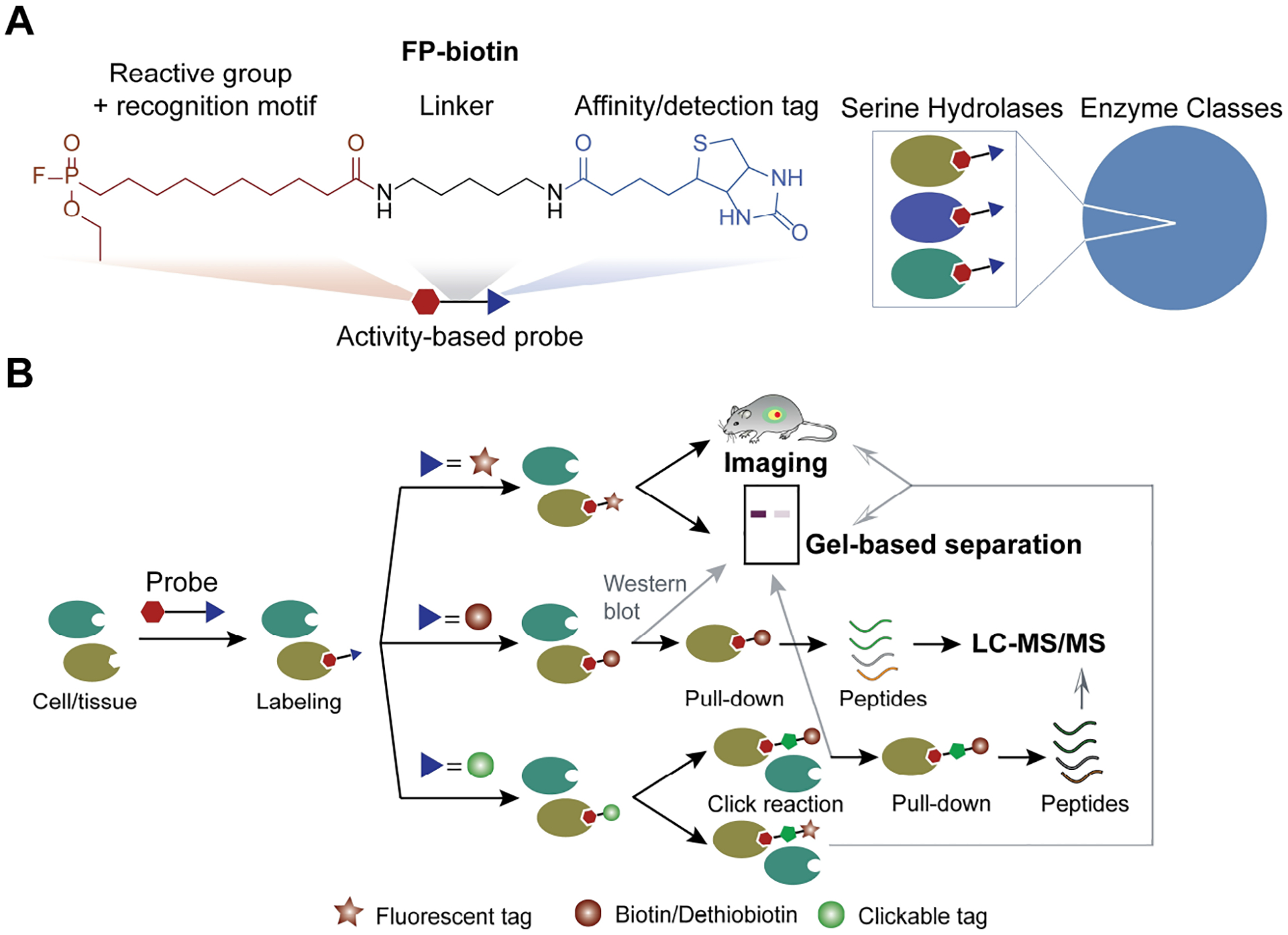
(A) Representative structure of the fluorophosphonate (FP) activity probe (ABP). In this example, the FP moiety serves as both enzyme recognition motif and covalent warhead. Integrated biotin tag provides a handle for biochemical pulldown as well as detection substrate. (B) General workflow for activity-based protein profiling (ABPP) of endogenous enzymes/proteins. The flexible ABPP platform supports multiple detection schemes.
In 2010, Cravatt extended the concept of activity-based probes to ‘reactivity-based’ probes (RBPs). Similar to ABPs, RBPs have a covalent warhead that targets a reactive moiety, typically an amino acid side chain in the cellular milieu, independent of protein or protein-family function. Electrophiles that target cysteine thiols groups[31] are the most common example, with the concept expanded to include methionine,[32] lysine,[33] tyrosine,[34] aspartate and glutamate,[35] as well as N-terminal glyoxylyl group.[36] As currently implemented, methods with RPBs are well-suited to identify the off-targets of covalent irreversible compounds.[36b,37] Cravatt recently extended the use of RBPs to characterize covalent reversible compounds as well.[38]
3. Competitive ABPP for Inhibitor Screening
In the past decades, competitive ABPP has emerged as a promising approach in early-stage drug discovery. As illustrated in Figure 2 (left), small molecules are co-incubated with the ABP to create a competitive binding assay.[6a] The ability to characterize small molecule activity against an enzyme class in native biological conditions can accelerate hit-to-lead and lead optimization.[6a] Since its initial introduction, the platform for competitive ABPP has been extended to encompass multiple detection strategies including optical, mass spectrometry, and qPCR.[6a,11,39] With limited throughput (compared to HTS), ABPP is not a practical strategy for screening diversity or other large compound collections; however recent applications demonstrate real promise for ABPP as a primary screen for focused libraries.[12f] We will discuss these strategies in detail and compare them with the corresponding strategies applied in an HTS manner.
Figure 2.
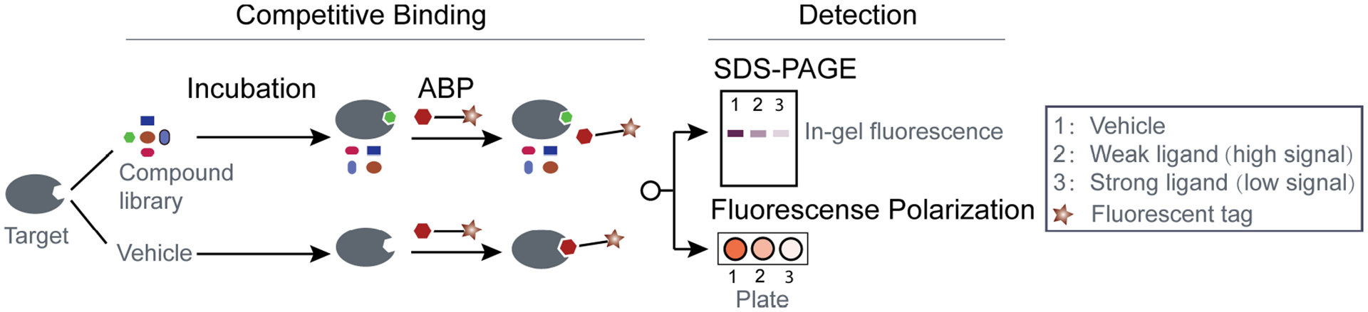
(left) General set-up for competitive ABPP comprising co-incubation of test compounds and activity-based probe (ABP). (right) Fluorescent based detection schemes to quantify test compound binding to target.
3.1. Chemiluminescence/Fluorescence Detection Based Competitive ABPP
Chemiluminescence provides sensitive and quantitative detection and was the readout originally employed in the first report of ABPP as part of a gel-based workflow.[9,40] For inhibitor screening in competitive ABPP, tissue or cell extract is preincubated with small molecules followed by co-incubation with fluorescent ABPs (Figure 2). Control and inhibitor-treated extracts are then resolved by SDS-PAGE, and the ABP-labeled enzymes are directly visualized by detection of in-gel fluorescence. Enzymes bound by inhibitors with increasing potency appear with reduced signal intensity as they are less available for conjugation with the ABP. Given the limited resolution of gel-based separation this approach is best suited for profiling modest numbers (~20) of probe-labeled protein targets. Even though the practical throughput of gel-based competitive ABPP (~100 runs) is much lower than HTS, it has nonetheless proven to be a robust approach. Successful applications of this workflow in inhibitor development span serine hydrolases, including fatty acid amide hydrolase,[41] monoacylglycerol lipase (MAGL),[42] diacylglycerol lipases,[43] KIAA1363,[12d] α/β hydrolase domain-containing protein,[44] and cysteine proteases.[12a,45]
Cravatt leveraged the concept of target-class ABP to create a hybrid ABPP-HTS platform (FluoPol-ABPP) suitable for screening understudied proteins. Poorly characterized proteins often lack validated substrates, making it difficult to develop an optimized biochemical assay for quantitative detection in HTS. The FluoPol-ABPP approach is based on detecting a change in fluorescence polarization when a fluorescent ABP binds the protein target (Figure 2). Small molecules compete with the ABP for target binding; test compounds that displaced the ABP lead to reduced polarization.[46] Using different fluorescent ABPs, including fluorophosphonate (FP)-rhodamine, F-amidine (RFA)-rhodamine, rhodamine-conjugated phenyl sulfonate ester, or a cyclophellitol aziridine probe with tetramethylrhodamine (TMR), inhibitor screening against serine hydrolase RBBP9,[46] protein arginine deiminases (PAD),[47] GSTO1,[48] and glucosylceramidase (GBA2)[49] has been successfully employed with libraries containing 350 to 300 K compounds.
3.2. Mass Spectrometry-Based Competitive ABPP
In much the same way that gel-free mass spectrometry approaches have replaced gel-based workflows, quantitative LC–MS/MS is gaining wide adoption as a direct detection scheme for competitive ABPP (LC–MS ABPP). As illustrated in Figure 3, test compounds and ABP are co-incubated and compete for binding to the proteins in the target-class of interest. To enable biochemical enrichment, the ABP is elaborated with a direct (e.g., biotin) or latent (e.g., alkyne) affinity handle. After pulldown, a number of proteomic workflows and quantitative mass spectrometry acquisition strategies are available to establish small molecule selectivity profiles across the enzyme family. Detailed comparisons of these different methods are beyond the scope of this review and are covered elsewhere.[50] Bypassing the gel provides a significant improvement in throughput while maintaining the advantage of selectivity assessment against enzymes in their native state. Compared to typical HTS approaches, the chemical space analyzed in LC–MS ABPP is relatively modest. However, because each compound is tested against many members of the enzyme family simultaneously, the total number of data points is still substantial. This high-content screen format enables rapid SAR assessment which can streamline subsequent chemical optimization of hit compounds. To date, LC–MS ABPP has been successfully used for development of both covalent and reversible inhibitors against multiple gene families, including kinases, deubiquitylating enzymes (DUBs), and fatty acid amide hydrolases.[12e,f, 51] Advances in separations and mass spectrometry technologies have improved throughput and quantitative performance in ABPP. For example, as originally described kinome ABPP, later commercialized as KiNativ, quantified compound binding to ~75 protein kinases in an approximate 2-hr analysis.[20] Recently, next-generation targeted MS/MS analysis (PRM-LIVE) demonstrated profiling depth of 220 endogenous kinases in a 60-min MS/MS acquisition.[52] Similar performance improvements have been achieved with ABPP for less well studied gene families. The family of deubiquitinating enzymes (DUBs) spans 100 proteins and can be profiled by use of a ubiquitin activity probe.[53] As with kinases, researchers continue to push the limits of coverage and throughput, with a recent example leveraging integrated preconcentration chip-based electrophoresis with targeted mass spectrometry to achieve profiling of ~50 endogenous DUBs in a rapid 15-min CE-MS/MS acquisition.[54]
Figure 3.
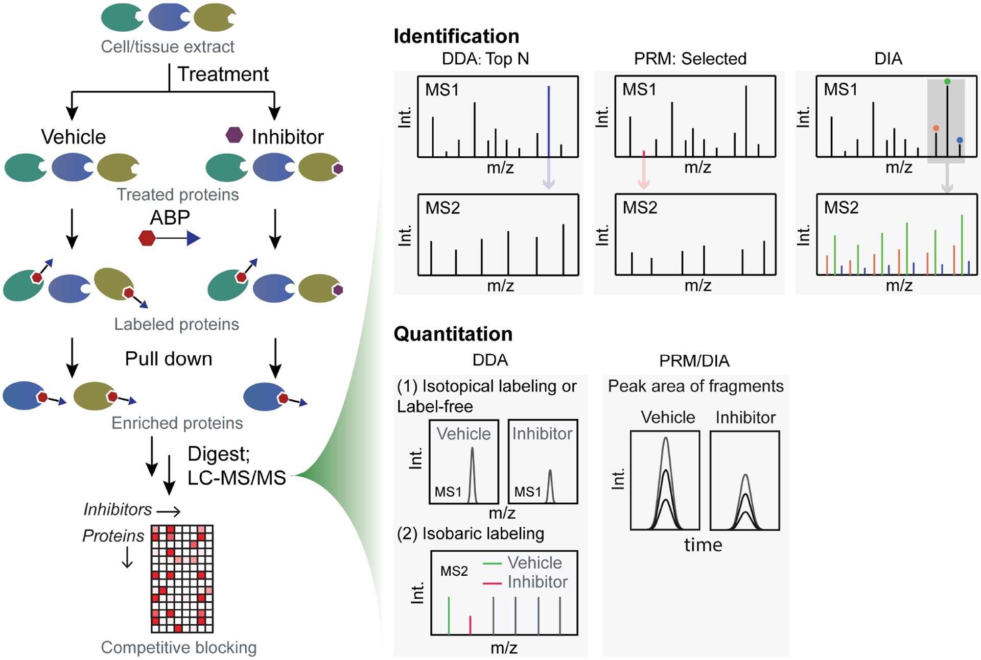
General workflow for LC–MS/MS-based competitive ABPP (LC–MS ABPP) as used to screen the binding behavior of small molecules against a target enzyme class. As reviewed (Ref. [50]), a variety of mass spectrometry data acquisition strategies, including data-dependent (DDA), data-independent (DIA) and parallel reaction monitoring (PRM), have been used for identification of ABP-bound enzymes and quantification of small molecule binding.
3.3. qPCR-Based Competitive ABPP
DNA-encoded libraries (DEL) span tremendous chemical diversity and can be used in very high throughput target-based screens; as a result, this approach is rapidly gaining momentum in small molecule drug discovery.[55] As shown in Figure 4A, DEL-based HTS usually starts with an immobilized protein target, followed by incubation with the DNA-encoded small molecule library,[56] a series of washing steps to remove unbound molecules, and finally elution of protein-bound ligands. Identifying bound ligands is performed by a combination of quantitative PCR (qPCR) followed by next-generation sequencing. Although only recently introduced, DEL screening has produced hits and leads that were ultimately developed into clinical candidates.[57]
Figure 4.
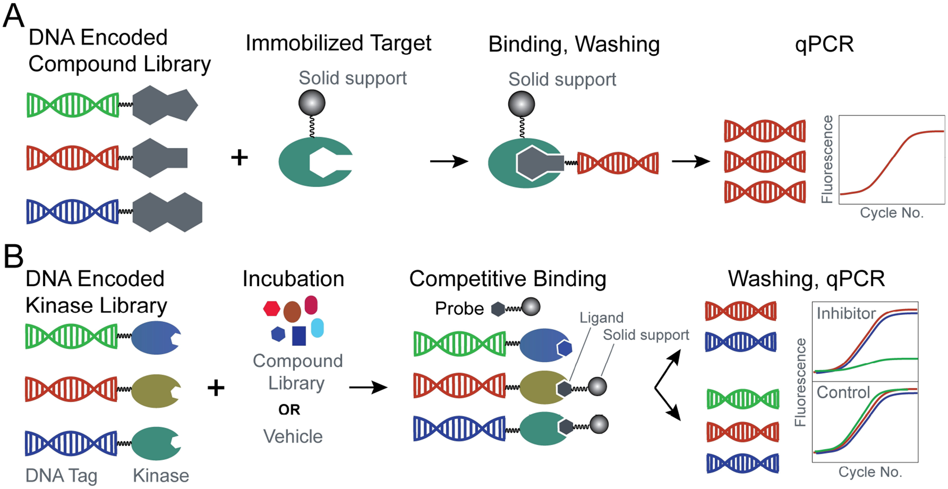
The concept of DNA-barcoding as used in high-throughput screening. (A) DNA-encoded small molecules are incubated with a purified target tethered to a bead or other solid support. qPCR is used to quantify small molecules bound to the target. (B) DNA-encoded kinases are incubated with test compounds and then co-incubated with ABPs tethered to beads. qPCR quantifies the relative enrichment of kinases on the beads as an indirect measure of small molecule binding.
In 2011, the concept of DEL-based HTS was incorporated in a competitive ABPP workflow to provide selectivity profiling of kinase inhibitors; this platform is available commercially as KINOMEscan (Figure 4B).[39] In contrast to most implementations of DEL screening, KINOMEscan utilizes a kinase library whereby each enzyme (full-length constructs for small, single-domain kinases, and catalytic domain constructs for multidomain kinases) is encoded with a unique DNA tag. A competitive binding assay is established by incubation of the tagged kinases with a test compound, followed by co-incubation with a solid-phase ABP comprising magnetic beads coated with a number of selective kinase inhibitors (similar to kinobeads or MIBs, see Section 2 above). A small molecule with high binding affinity for one or more tagged kinase(s) will prevent the interaction of these targets with the ABP. After capture of the magnetic beads, test compound selectivity is assessed by qPCR to quantify kinases that remain bound to the solid phase ABP. This platform has been widely used for the development of kinase inhibitors.[55b,58]
3.4. ABPP as a Primary Screen for Focused Libraries
Target-based screening has proven successful in drug discovery,[59] with related innovations such as DEL allowing interrogation of ever-larger chemical space. However, most HTS campaigns are resource intensive and largely inaccessible for all but the largest R&D organizations. Fortunately, increased availability of experimentally determined (Structural Genomics Consortium[60]) or computationally predicted (alpha-fold[61]) protein structures, combined with docking software[62] and improvements in virtual screening[63] have begun to democratize the process of building focused libraries for targets or gene families of interest. A comprehensive description of the components comprising virtual screening and molecular docking are outside the scope of this review and are detailed elsewhere.[64] Of particular note for integration with ABPP, these computational tools provide a myriad of bootstrapping[65] or post-processing filters.[66] While still requiring a degree of specialized expertise, virtual screening nonetheless holds great potential as a robust, fast, cost-effective, and hence widely accessible approach that often yields a modest-sized, focused compound library well-suited for ABPP (Figure 5). In a recent study, researchers used virtual screening (ZINC and MCE compound libraries) to analyze an input set of >10 million compounds to yield a refined library of ~400 potential binders against mtbDHFR. They then used hybrid FluoPol-ABPP to identify and further characterize one hit (AF-353) as a potent mtbDHFR inhibitor.[67] In fact, this compound exhibited 50-fold selectivity for tuberculosis DHFR versus the human protein, demonstrating how the combination of computational and biochemical approaches can streamline identification of lead compounds with favorable therapeutic windows.[67] Virtual screening has also been utilized to prioritize promising candidates in irreversible libraries. To develop selective inhibitors of the MAP2 kinase, MKK7, Nir London’s group used their DOCKovalent software to select 10 acrylamide test compounds from a larger library of ~120,000 electrophilic fragments. In silico docking analysis was used to guide design of analogs which were tested for kinase selectivity with gel-based ABPP.[68]
Figure 5.
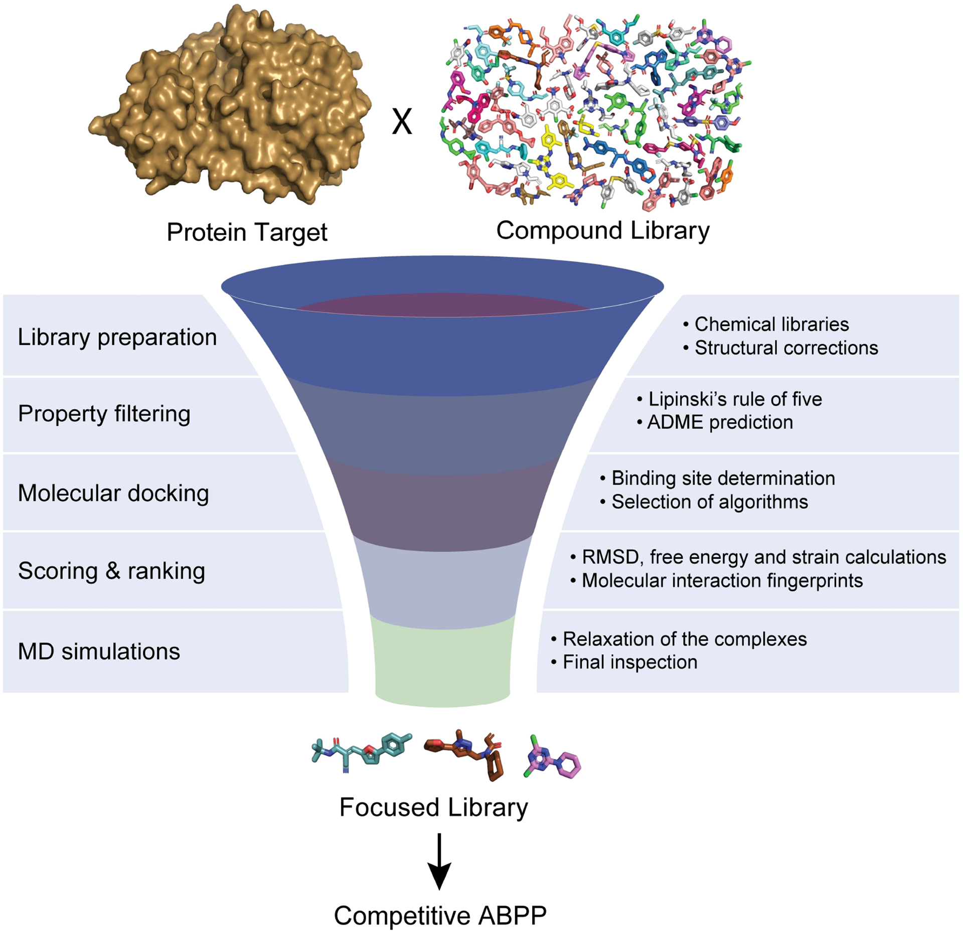
Workflow for in silico, structure-guided virtual screening
ABPP is also finding wider use to screen existing focused libraries. For example, Baggelaar and colleagues developed a new lactam-based ABP and found that it could bind a subset of serine hydrolases that were not addressed by existing organophosphate probes.[44e] The authors used their new ABP to screen a focused library of ~200 lipase-targeted compounds to identify an inhibitor of endogenous ABHD2, with potential applications in male contraceptives. In another study, also focused on serine hydrolases, Deng and co-workers utilized gel-based ABPP as part of a larger screening effort to identify reversible inhibitors against MAGL.[69] Here their focused library comprised ~300 commercially available natural products, chosen to bias the analysis towards new chemotypes for further development.
4. Case Studies
In this section we detail several examples in which ABPP played a significant role in the development and characterization of selective inhibitors, clinical candidates, or drugs.
4.1. MAGL Inhibitor
ABX-1431 is a selective CNS-penetrant monoacylglycerol lipase (MAGL) inhibitor.[13b] It was discovered and optimized through a structure-guided approach and then extensively evaluated by ABPP. In a series of intensive in vitro and in vivo gel-based competitive ABPP experiments, the compound was found to be potent and selective in human cell lines as well as prefrontal cortex homogenates prepared post-mortem from mouse, rat, dog, and human brains. Of note, in the human brain ABX-1431 inhibited MAGL with high selectively compared to 37 other serine hydrolases. Similar experiments in mice confirmed MAGL as the primary target, but also identified the serine hydrolase ABHD6 as one off-target. The authors then used a clickable analog of ABX-1431 in in vivo experiments to confirm selectivity across the proteome. In 2018, ABX-1431 entered a phase 2 clinical trial for the treatment of Tourette syndrome. The trial ended in 2020 due to the weak efficacy of ABX-1431 in suppressing motor tics.[70] However, in the interim researchers explored other indications for inhibition of MAGL and a new phase I trial was approved in 2021 to evaluate ABX-1431 for treatment of multiple sclerosis (ClinicalTrials.gov Identifier: NCT04990219). This example illustrates the effective use of gel-based competitive ABPP to assess target engagement and compound selectivity during inhibitor development.
4.2. First-In-Class KRASG12 C Drugs
KRAS is the most frequently mutated gene across all cancers with high penetrance in colorectal, lung, and pancreatic tumors.[71] KRAS had been considered ‘undruggable’ due to a paucity of binding pockets suitable for engagement by small molecules.[72] However, the G12C KRAS mutant presented a unique opportunity due to the presence of the mutant-specific cysteine thiol. In 2013, the Shokat group discovered the first covalent small molecule to selectively bind the KRAS(G12C)-GDP complex.[73] Building on this initial work, further structural optimization led to the development of ARS-853, ARS-1620, and finally the clinical KRASG12 C inhibitors including sotorasib (AMG510) and adagrasib (MRTX849).[37] AMG510 exemplifies the use of reactivity-based probes in an ABPP format for drug development. Specifically, the optimization of ARS-107 relied heavily on use of cysteine-directed RBPs to identify off-targets across the proteome. These studies progressed to ARS-1620 which had improved selectivity, but still bound one major off-target as identified by cysteine profiling.[37b] Interestingly as part of an atropisomer pair, ARS-1620 binds KRASG12C while the R-atropisomer is inactive.[37b,74] Further medicinal chemistry resulted in AMG510, which showed enhanced target engagement with KRASG12C, along with exquisite selectivity, binding only Cys-12 on the mutant out of >6,000 cysteines in across the proteome.[37c] In 2021, AMG510 was approved by the FDA for clinical use in lung cancer, becoming the first covalent drug against KRASG12C. This example demonstrates the utility of RBPs used in an ABPP format to support development of new covalent drugs.
4.3. Inhibitors Against DUB Family Enzymes
Deubiquitinating enzymes (DUBs) are an understudied class of ~100 proteases that cleave ubiquitin from protein substrates to regulate many cellular processes.[75] Members of the family have been linked to human cancer and neurodegenerative disease,[76] suggesting that DUB inhibitors could have a significant therapeutic impact. However, a lack of potent and selective chemical probes represents a significant hurdle in interrogating DUB biology or validating their potential as drug targets.[53,76]
VLX1570 was the first DUB inhibitor to enter phase II clinical trials (ClinicalTrials.gov Identifier: NCT02372240). It was reported to covalently bind and inhibit activity of the DUB USP14 by the Linder group in 2016.[77] Due to dose-limiting toxicity, the clinical trial for VLX1570 was terminated in 2018 (ClinicalTrials.gov Identifier: NCT02372240). Interestingly, VLX1570 possesses more than one Michael acceptor. In 2020, Tate and co-workers used a combination of western blot and ABPP to identify CIAPIN1 as one of the major off targets of VLX1570. The investigators hypothesized that covalent modification of CIAPIN1 could induce protein aggregations and cell death, providing a possible mechanism for toxicity associated with VLX1570.[78] This example illustrates how use of ABPP to characterize the off-targets of an advanced small molecule inhibitor could inform future optimization studies.
DUBs engage their cognate ubiquitin ligand through a myriad of interactions. We recently undertook a structure-guided approach to create a novel covalent library diversified against discrete regions in and around the catalytic site. As shown in Figure 6, we paired our focused library of ~180 covalent compounds with a ubiquitin ABP and LC–MS ABPP to obtain hits against 56 endogenous DUBs.[12f] Among the 45 hits, we identified a novel azetidine compound (WH-9943–103 C) that was potent (<1 μM) and selective for the understudied DUB, VCPIP1. Importantly the use of LC–MS ABPP as a primary screen platform enabled us to perform SAR deconvolution for potency and selectivity simultaneously; these results guided the development of a 20-compound expansion set that rapidly yielded a first-in-class, selective 70 nM covalent probe for VCPIP1. This study provides a road map for the use of LC–MS based ABPP to accelerate systematic development of selective chemical probes for understudied gene families.
Figure 6.
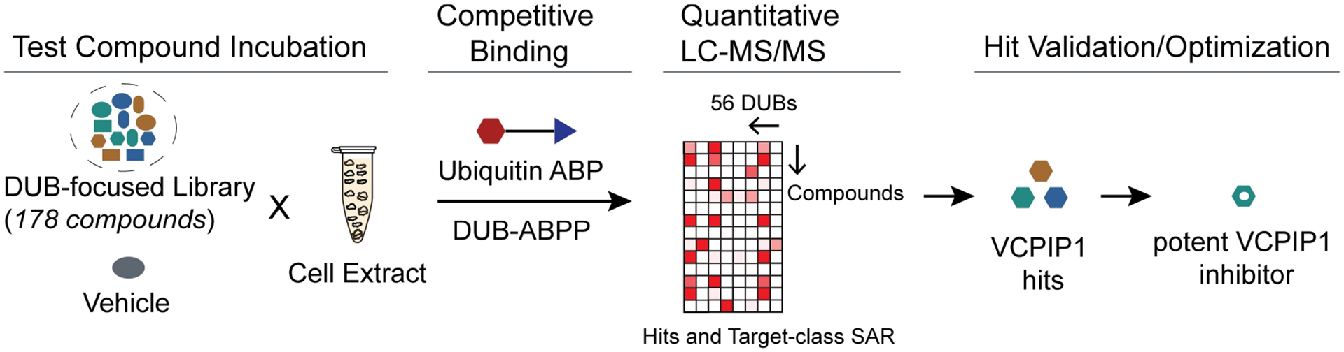
Workflow for DUB ABPP using a focused covalent library paired with LC–MS/MS. Reproducible detection of 56 endogenous DUBs across multiple LC–MS/MS experiments enabled target-class SAR and development of first-in-class covalent inhibitor of the understudied DUB VCPIP1.
4.4. Mycobacterium Tuberculosis (Mtb) Inhibitor
The proliferation of multi-antibiotic resistance bacteria represents an area in need of improved screening to streamline drug discovery.[79] Historically, researchers have relied heavily on phenotypic screening to identify new antibiotics; however, this strategy provides limited understanding of the mechanism of action of hit compounds. In principle coupling high throughput phenotypic screening with ABPP streamlines target deconvolution and validation.[80] In a recent example, two groups[14a,80] independently discovered novel chemotypes, 1,2,3-triazole urea and 7-urea chloroisocoumarin, that inhibit Mtb growth through disruption of cell wall biosynthesis. Both groups used competitive ABPP to identify members of the serine hydrolase family as potential targets. Li et al. focused on 1,2,3-triazole urea compounds, using active (AA692) and inactive (AA702) isomers of a hit series to narrow their target set to 5 serine hydrolases. Babin et al. used a 7-urea chloroisocoumarin chemotype, exemplified by JCP276, to identify 9 hydrolase targets. Interestingly, disruption of these hydrolases individually resulted in Mtb growth defects, suggesting productive polypharmacology for JCP276.[80]
4.5. Optimization of BTK Drugs
The human kinome comprises more than 500 enzymes many of which are well-validated as human disease targets.[81] As of 2021 there were 68 FDA-approved drugs targeting kinases.[82] ABPP has a strong history in enabling kinase drug discovery. The Bruton’s tyrosine kinase (BTK) inhibitor PCI-32765 was originally developed through a collaboration between Celera and Pharmacyclics.[83] In 2013 the FDA approved this covalent drug, ‘Ibrutinib’, for mantel cell lymphoma; use for chronic lymphocytic leukemia (CLL) and Waldenström’s macroglobulinemia were approved in 2014 and 2015, respectively.[84] In 2014, Cravatt et al. utilized a combination of gel- and LC–MS/MS-based ABPP to investigate the landscape of on- and off-targets as a function of Ibrutinib concentration. Based on these data they undertook a modest chemical optimization campaign combined with competitive ABPP to discover that methylation of the terminal Michael acceptor of ibrutinib did not appreciably diminish potency against BTK but did reduce the number of off-targets in the proteome.[51d] Driven by the clinical success of Ibrutinib, other structurally related covalent drugs including acalabrutinib and zanubrutinib, also with high potency and exquisite selectivity, have been approved primarily for cancer therapy.[85] This example illustrates the utility of ABPP to guide improvements in existing drugs.
5. Summary and Outlook
Activity-based protein profiling has become a foundational technology for identifying ligands to proteins, highlighting potential off-targets, characterizing new binding pockets, and finding new chemical starting points for clinical drugs.[1b,86] The general concept has proven amenable to innovations in terms of sample preparation and automation, chemistry for new activity probes and reporters, as well as improved quantification and diversified detection schemes. Given the flexibility and power of the platform, it is not surprising that ABPP is seeing widespread adoption by the research community, with a significant upward trend in PubMed hits. The radar plots below (Figure 7) illustrate the relative strengths of the approaches reviewed herein. Going forward, continued development of activity- and reactivity-based probes will broaden the addressable landscape of human proteins amenable to characterization by ABPP. Further developments in reporter probes as well as mass spectrometry and other detector technologies will support improved throughput. Similarly, advances in computational screening and virtual hit prioritization may yield rapid assembly of focused libraries that are compatible with current throughput of ABPP platforms. As highlighted in the Case Studies, strategic use of ABPP promises to accelerate drug discovery for human disease.
Figure 7.
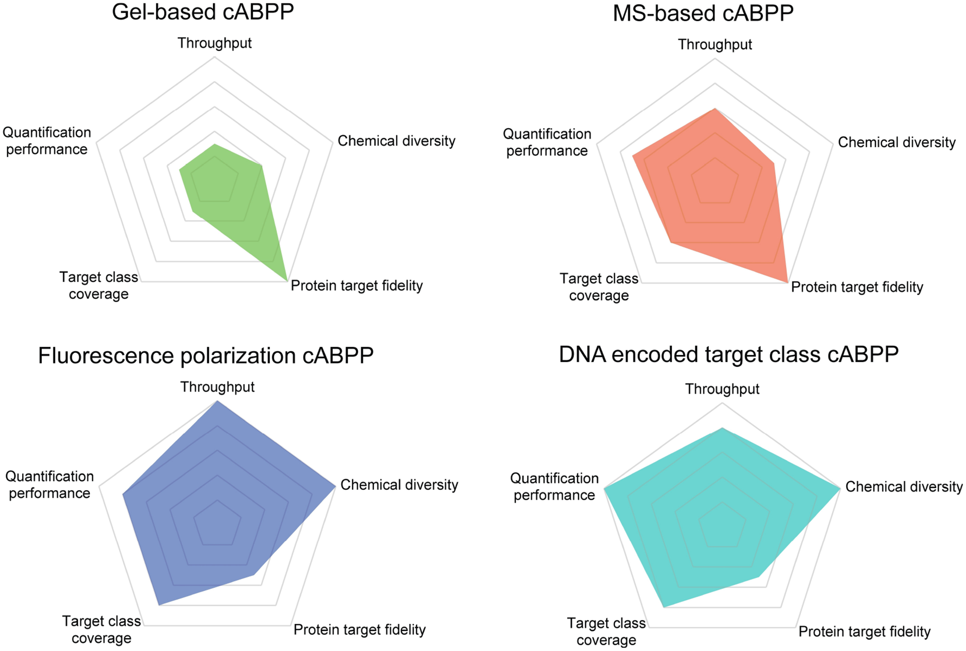
Radar plots illustrate the relative strengths of different strategies used in competitive ABPP (cABPP). The estimation of performance metrics is based on single assay.
Acknowledgements
The authors acknowledge generous support from National Cancer Center (to H.Z.), The Mark Foundation for Cancer Research (to S.J.B. and J.A.M.), Ellison Foundation (to S.J.B.), the National Institutes of Health (CA233800 and CA247671 to S.J.B. and J.A.M., U24DK116204 to J.A.M.), and the Massachusetts Life Science Center (to S.J.B. and J.A.M.).
Competing Financial Interests
J.A.M. is a founder, equity holder, and advisor to Entact Bio, serves on the SAB of 908 Devices, and receives sponsored research funding from Vertex, AstraZeneca, Taiho, Spring-works and TUO Therapeutics. S.J.B. is a founder, equity holder, and advisor to Entact Bio and serves on the SAB of Adenoid Cystic Carcinoma Foundation. S.J.B. currently receives funding from AbbVie, Tuo Therapeutics, and Kinogen, and has received in-kind services from AbbVie and Novartis Institutes for Biomedical Research during the past 24 months. The remaining authors declare no competing interests.
Biographies

He (Eric) Zhu obtained a PhD in analytical chemistry from Georgia State University and an MS in medicinal chemistry from Ocean University of China. He is currently a National Cancer Center research fellow in the Marto Lab at the Dana-Farber Cancer Institute. Dr. Zhu’s research primarily focuses on developing innovative mass spectrometry strategies to characterize protein-compound interactions and quantify the dynamics of protein post-translational modifications.

Mona Sharafi received her PhD from University of Vermont where she focused on synthesis and modeling of selective supramolecular catalysts. She is currently a postdoctoral research fellow in the Buhrlage lab at Dana Farber Cancer Institute working on development of potent and selective inhibitors targeting deubiquitinating enzymes by combining her skills in synthetic and computational chemistry.

Wei Pin Teh received his PhD in Chemistry, focusing on synthetic organic methodology from University of Washington with Prof. Forrest Michael. He is currently a postdoctoral research fellow in the Buhrlage lab at Dana-Farber Cancer Institute in Boston. His current research focuses on developing bivalent compounds targeting deubiquitinating enzymes (DUB) and chemical tools to understand the role of DUBs in biological systems.

Ariana Bratt is a graduate student in the Chemical Biology PhD program at Harvard in Sara Buhrlage’s lab at the Dana-Farber Cancer Institute. Her research involves utilizing chemical biology tools to study the biology of deubiquitinating enzymes and their role in disease. She received her BS in chemistry from Rice University and MD from UCSF.

Sara Buhrlage, PhD is an Associate Professor holding joint appointments in the Dana-Farber Cancer Institute department of Cancer Biology and the Harvard Medical School department of Biological Chemistry and Molecular Pharmacology. Dr. Buhrlage’s research is focused on development of new chemical probes and therapeutic strategies for deubiquitinases (DUBs). Her research has provided new insights into the chemical tractability of DUBs, approaches for illuminating DUB function and therapeutic potential and roles of DUBs in cancer. Dr. Buhrlage earned a Doctor of Philosophy in organic chemistry from the University of Michigan and completed a postdoctoral fellowship at the Broad Institute.

Jarrod Marto, Ph.D. joined the Dana-Farber Cancer Institute in 2004 where he is currently an Associate Professor in the Departments of Cancer Biology and Oncologic Pathology, with appointments in the Department of Pathology at the Brigham and Women’s Hospital and Harvard Medical School. Dr. Marto uses mass spectrometry as a primary discovery tool to understand how genomic alterations, environmental insults, or the action of small molecule chemical probes impact the functional proteome, including protein post-translational modifications, biochemical complexes, signaling pathways, or how they manifest en masse as phenotypic or disease signatures. He earned his Ph.D. in analytical chemistry at The Ohio State University with Alan Marshall and went on to postdoctoral studies at the University of Virginia with Don Hunt.
References
- [1].a) Blagg J, Workman P, Cancer Cell 2017, 32, 9–25; [DOI] [PMC free article] [PubMed] [Google Scholar]; b) Boike L, Henning NJ, Nomura DK, Nat. Rev. Drug Discovery 2022, 21, 881–898; [DOI] [PMC free article] [PubMed] [Google Scholar]; c) Frye SV, Nat. Chem. Biol 2010, 6, 159–161. [DOI] [PubMed] [Google Scholar]
- [2].a) Cohen P, Cross D, Janne PA, Nat. Rev. Drug Discovery 2021, 20, 551–569; [DOI] [PMC free article] [PubMed] [Google Scholar]; b) Wang Y, Zhang S, Li F, Zhou Y, Zhang Y, Wang Z, Zhang R, Zhu J, Ren Y, Tan Y, Qin C, Li Y, Li X, Chen Y, Zhu F, Nucleic Acids Res. 2020, 48, D1031–D1041. [DOI] [PMC free article] [PubMed] [Google Scholar]
- [3].a) Eisenberg D, Marcotte EM, Xenarios I, Yeates TO, Nature 2000, 405, 823–826; [DOI] [PubMed] [Google Scholar]; b) Cruz LM, Trefflich S, Weiss VA, Castro MAA, Methods Mol. Biol 2017, 1654, 55–75. [DOI] [PubMed] [Google Scholar]
- [4].Workman P, Collins I, Chem. Biol 2010, 17, 561–577. [DOI] [PMC free article] [PubMed] [Google Scholar]
- [5].Wright MH, Sieber SA, Nat. Prod. Rep 2016, 33, 681–708. [DOI] [PMC free article] [PubMed] [Google Scholar]
- [6].a) Niphakis MJ, Cravatt BF, Annu. Rev. Biochem 2014, 83, 341–377; [DOI] [PubMed] [Google Scholar]; b) Bunnage ME, Chekler EL, Jones LH, Nat. Chem. Biol 2013, 9, 195–199. [DOI] [PubMed] [Google Scholar]
- [7].Jia Y, Yun CH, Park E, Ercan D, Manuia M, Juarez J, Xu C, Rhee K, Chen T, Zhang H, Palakurthi S, Jang J, Lelais G, DiDonato M, Bursulaya B, Michellys PY, Epple R, Marsilje TH, McNeill M, Lu W, Harris J, Bender S, Wong KK, Janne PA, Eck MJ, Nature 2016, 534, 129–132. [DOI] [PMC free article] [PubMed] [Google Scholar]
- [8].Sams–Dodd F, Drug Discovery Today 2005, 10, 139–147. [DOI] [PubMed] [Google Scholar]
- [9].Liu Y, Patricelli MP, Cravatt BF, Proc. Natl. Acad. Sci. USA 1999, 96, 14694–14699. [DOI] [PMC free article] [PubMed] [Google Scholar]
- [10].Adibekian A, Martin BR, Wang C, Hsu KL, Bachovchin DA, Niessen S, Hoover H, Cravatt BF, Nat. Chem. Biol 2011, 7, 469–478. [DOI] [PMC free article] [PubMed] [Google Scholar]
- [11].Carvalho LAR, Bernardes GJL, ChemMedChem. 2022, 17, e202200174. [DOI] [PMC free article] [PubMed] [Google Scholar]
- [12].a) Berger AB, Witte MD, Denault JB, Sadaghiani AM, Sexton KM, Salvesen GS, Bogyo M, Mol. Cell 2006, 23, 509–521; [DOI] [PubMed] [Google Scholar]; b) Rolen U, Kobzeva V, Gasparjan N, Ovaa H, Winberg G, Kisseljov F, Masucci MG, Mol. Carcinog 2006, 45, 260–269; [DOI] [PubMed] [Google Scholar]; c) Salisbury CM, Cravatt BF, J. Am. Chem. Soc 2008, 130, 2184–2194; [DOI] [PubMed] [Google Scholar]; d) Chang JW, Nomura DK, Cravatt BF, Chem. Biol 2011, 18, 476–484; [DOI] [PMC free article] [PubMed] [Google Scholar]; e) van Esbroeck ACM, Janssen APA, Cognetta AB 3rd, Ogasawara D, Shpak G, van der Kroeg M, Kantae V, Baggelaar MP, de Vrij FMS, Deng H, Allara M, Fezza F, Lin Z, van der Wel T, Soethoudt M, Mock ED, den Dulk H, Baak IL, Florea BI, Hendriks G, De Petrocellis L, Overkleeft HS, Hankemeier T, De Zeeuw CI, Di Marzo V, Maccarrone M, Cravatt BF, Kushner SA, van der Stelt M, Science 2017, 356, 1084–1087; [DOI] [PMC free article] [PubMed] [Google Scholar]; f) Chan WC, Liu X, Magin RS, Girardi NM, Ficarro SB, Hu W, Tarazona Guzman MI, Starnbach CA, Felix A, Adelmant G, Varca AC, Hu B, Bratt AS, DaSilva E, Schauer NJ, Jaen Maisonet I, Dolen EK, Ayala AX, Marto JA, Buhrlage SJ, Nat. Commun 2023, 14, 686. [DOI] [PMC free article] [PubMed] [Google Scholar]
- [13].a) Janssen APA, van der Vliet D, Bakker AT, Jiang M, Grimm SH, Campiani G, Butini S, van der Stelt M, ACS Chem. Biol 2018, 13, 2406–2413; [DOI] [PMC free article] [PubMed] [Google Scholar]; b) Cisar JS, Weber OD, Clapper JR, Blankman JL, Henry CL, Simon GM, Alexander JP, Jones TK, Ezekowitz RAB, O’Neill GP, Grice CA, J. Med. Chem 2018, 61, 9062–9084. [DOI] [PubMed] [Google Scholar]
- [14].a) Li M, Patel HV, Cognetta AB 3rd, Smith TC 2nd, Mallick I, Cavalier JF, Previti ML, Canaan S, Aldridge BB, Cravatt BF, Seeliger JC, Cell Chem. Biol 2022, 29, 883–896.e5; [DOI] [PMC free article] [PubMed] [Google Scholar]; b) Prothiwa M, Englmaier F, Bottcher T, J. Am. Chem. Soc 2018, 140, 14019–14023; [DOI] [PubMed] [Google Scholar]; c) Schmidt C, Zollo M, Bonsignore R, Casini A, Hacker SM, Chem. Commun. (Camb.) 2022, 58, 5526–5529; [DOI] [PubMed] [Google Scholar]; d) Hoegl A, Nodwell MB, Kirsch VC, Bach NC, Pfanzelt M, Stahl M, Schneider S, Sieber SA, Nat. Chem 2018, 10, 1234–1245; [DOI] [PMC free article] [PubMed] [Google Scholar]; e) Sharifzadeh S, Boersma MJ, Kocaoglu O, Shokri A, Brown CL, Shirley JD, Winkler ME, Carlson EE, ACS Chem. Biol 2017, 12, 2849–2857. [DOI] [PMC free article] [PubMed] [Google Scholar]
- [15].Penalver L, Schmid P, Szamosvari D, Schildknecht S, Globisch C, Sawade K, Peter C, Bottcher T, Angew. Chem. Int. Ed. Engl 2021, 60, 6799–6806. [DOI] [PMC free article] [PubMed] [Google Scholar]
- [16].Benns HJ, Wincott CJ, Tate EW, Child MA, Curr. Opin. Chem. Biol 2021, 60, 20–29. [DOI] [PubMed] [Google Scholar]
- [17].Greenbaum D, Medzihradszky KF, Burlingame A, Bogyo M, Chem. Biol 2000, 7, 569–581. [DOI] [PubMed] [Google Scholar]
- [18].Saghatelian A, Jessani N, Joseph A, Humphrey M, Cravatt BF, Proc. Natl. Acad. Sci. USA 2004, 101, 10000–10005. [DOI] [PMC free article] [PubMed] [Google Scholar]
- [19].Kumar S, Zhou B, Liang F, Wang WQ, Huang Z, Zhang ZY, Proc. Natl. Acad. Sci. USA 2004, 101, 7943–7948. [DOI] [PMC free article] [PubMed] [Google Scholar]
- [20].Patricelli MP, Szardenings AK, Liyanage M, Nomanbhoy TK, Wu M, Weissig H, Aban A, Chun D, Tanner S, Kozarich JW, Biochemistry 2007, 46, 350–358. [DOI] [PubMed] [Google Scholar]
- [21].a) Vocadlo DJ, Bertozzi CR, Angew. Chem. Int. Ed. Engl 2004, 43, 5338–5342; [DOI] [PubMed] [Google Scholar]; b) Stubbs KA, Scaffidi A, Debowski AW, Mark BL, Stick RV, Vocadlo DJ, J. Am. Chem. Soc 2008, 130, 327–335. [DOI] [PubMed] [Google Scholar]
- [22].Krysiak JM, Kreuzer J, Macheroux P, Hermetter A, Sieber SA, Breinbauer R, Angew. Chem. Int. Ed. Engl 2012, 51, 7035–7040. [DOI] [PMC free article] [PubMed] [Google Scholar]
- [23].Kahler JP, Verhelst SHL, RSC Chem. Biol 2021, 2, 1285–1290. [DOI] [PMC free article] [PubMed] [Google Scholar]
- [24].a) Dubinsky L, Jarosz LM, Amara N, Krief P, Kravchenko VV, Krom BP, Meijler MM, Chem. Commun. (Camb.) 2009, 7378–7380; [DOI] [PubMed] [Google Scholar]; b) Ranjitkar P, Perera BG, Swaney DL, Hari SB, Larson ET, Krishnamurty R, Merritt EA, Villen J, Maly DJ, J. Am. Chem. Soc 2012, 134, 19017–19025; [DOI] [PMC free article] [PubMed] [Google Scholar]; c) Hulce JJ, Cognetta AB, Niphakis MJ, Tully SE, Cravatt BF, Nat. Methods 2013, 10, 259–264; [DOI] [PMC free article] [PubMed] [Google Scholar]; d) Parker CG, Galmozzi A, Wang Y, Correia BE, Sasaki K, Joslyn CM, Kim AS, Cavallaro CL, Lawrence RM, Johnson SR, Narvaiza I, Saez E, Cravatt BF, Cell 2017, 168, 527–541.e5. [DOI] [PMC free article] [PubMed] [Google Scholar]
- [25].a) Ekkebus R, van Kasteren SI, Kulathu Y, Scholten A, Berlin I, Geurink PP, de Jong A, Goerdayal S, Neefjes J, Heck AJ, Komander D, Ovaa H, J. Am. Chem. Soc 2013, 135, 2867–2870; [DOI] [PMC free article] [PubMed] [Google Scholar]; b) de Jong A, Merkx R, Berlin I, Rodenko B, Wijdeven RH, El Atmioui D, Yalcin Z, Robson CN, Neefjes JJ, Ovaa H, ChemBioChem 2012, 13, 2251–2258. [DOI] [PMC free article] [PubMed] [Google Scholar]
- [26].Cravatt BF, Wright AT, Kozarich JW, Annu. Rev. Biochem 2008, 77, 383–414. [DOI] [PubMed] [Google Scholar]
- [27].a) Blum G, Mullins SR, Keren K, Fonovic M, Jedeszko C, Rice MJ, Sloane BF, Bogyo M, Nat. Chem. Biol 2005, 1, 203–209; [DOI] [PubMed] [Google Scholar]; b) Blum G, von Degenfeld G, Merchant MJ, Blau HM, Bogyo M, Nat. Chem. Biol 2007, 3, 668–677; [DOI] [PubMed] [Google Scholar]; c) Edgington LE, Berger AB, Blum G, Albrow VE, Paulick MG, Lineberry N, Bogyo M, Nat. Med 2009, 15, 967–973; [DOI] [PMC free article] [PubMed] [Google Scholar]; d) Verdoes M, Oresic Bender K, Segal E, van der Linden WA, Syed S, Withana NP, Sanman LE, Bogyo M, J. Am. Chem. Soc 2013, 135, 14726–14730. [DOI] [PMC free article] [PubMed] [Google Scholar]
- [28].Yu WL, Guizzunti G, Foley TL, Burkart MD, La Clair JJ, J. Nat. Prod 2010, 73, 1659–1666. [DOI] [PubMed] [Google Scholar]
- [29].a) Bantscheff M, Eberhard D, Abraham Y, Bastuck S, Boesche M, Hobson S, Mathieson T, Perrin J, Raida M, Rau C, Reader V, Sweetman G, Bauer A, Bouwmeester T, Hopf C, Kruse U, Neubauer G, Ramsden N, Rick J, Kuster B, Drewes G, Nat. Biotechnol 2007, 25, 1035–1044; [DOI] [PubMed] [Google Scholar]; b) Duncan JS, Whittle MC, Nakamura K, Abell AN, Midland AA, Zawistowski JS, Johnson NL, Granger DA, Jordan NV, Darr DB, Usary J, Kuan PF, Smalley DM, Major B, He X, Hoadley KA, Zhou B, Sharpless NE, Perou CM, Kim WY, Gomez SM, Chen X, Jin J, Frye SV, Earp HS, Graves LM, Johnson GL, Cell 2012, 149, 307–321. [DOI] [PMC free article] [PubMed] [Google Scholar]
- [30].a) Fang H, Peng B, Ong SY, Wu Q, Li L, Yao SQ, Chem. Sci 2021, 12, 8288–8310; [DOI] [PMC free article] [PubMed] [Google Scholar]; b) Wang S, Tian, Wang M, Wang M, Sun GB, Sun XB, Front. Pharmacol 2018, 9, 353. [DOI] [PMC free article] [PubMed] [Google Scholar]
- [31].Weerapana E, Wang C, Simon GM, Richter F, Khare S, Dillon MB, Bachovchin DA, Mowen K, Baker D, Cravatt BF, Nature 2010, 468, 790–795. [DOI] [PMC free article] [PubMed] [Google Scholar]
- [32].Lin S, Yang X, Jia S, Weeks AM, Hornsby M, Lee PS, Nichiporuk RV, Iavarone AT, Wells JA, Toste FD, Chang CJ, Science 2017, 355, 597–602. [DOI] [PMC free article] [PubMed] [Google Scholar]
- [33].Hacker SM, Backus KM, Lazear MR, Forli S, Correia BE, Cravatt BF, Nat. Chem 2017, 9, 1181–1190. [DOI] [PMC free article] [PubMed] [Google Scholar]
- [34].Hahm HS, Toroitich EK, Borne AL, Brulet JW, Libby AH, Yuan K, Ware TB, McCloud RL, Ciancone AM, Hsu KL, Nat. Chem. Biol 2020, 16, 150–159. [DOI] [PMC free article] [PubMed] [Google Scholar]
- [35].a) Bach K, Beerkens BLH, Zanon PRA, Hacker SM, ACS Cent. Sci 2020, 6, 546–554; [DOI] [PMC free article] [PubMed] [Google Scholar]; b) Ma N, Hu J, Zhang ZM, Liu W, Huang M, Fan Y, Yin X, Wang J, Ding K, Ye W, Li Z, J. Am. Chem. Soc 2020, 142, 6051–6059. [DOI] [PubMed] [Google Scholar]
- [36].a) Matthews ML, He L, Horning BD, Olson EJ, Correia BE, Yates JR 3rd, Dawson PE, Cravatt BF, Nat. Chem 2017, 9, 234–243; [DOI] [PMC free article] [PubMed] [Google Scholar]; b) Wang X, Lin Z, Bustin KA, McKnight NR, Parsons WH, Matthews ML, J. Am. Chem. Soc 2022, 144, 5377–5388. [DOI] [PMC free article] [PubMed] [Google Scholar]
- [37].a) Patricelli MP, Janes MR, Li LS, Hansen R, Peters U, Kessler LV, Chen Y, Kucharski JM, Feng J, Ely T, Chen JH, Firdaus SJ, Babbar A, Ren P, Liu Y, Cancer Discov. 2016, 6, 316–329; [DOI] [PubMed] [Google Scholar]; b) Janes MR, Zhang J, Li LS, Hansen R, Peters U, Guo X, Chen Y, Babbar A, Firdaus SJ, Darjania L, Feng J, Chen JH, Li S, Li S, Long YO, Thach C, Liu Y, Zarieh A, Ely T, Kucharski JM, Kessler LV, Wu T, Yu K, Wang Y, Yao Y, Deng X, Zarrinkar PP, Brehmer D, Dhanak D, Lorenzi MV, Hu–Lowe D, Patricelli MP, Ren P, Liu Y, Cell 2018, 172, 578–589.e17; [DOI] [PubMed] [Google Scholar]; c) Canon J, Rex K, Saiki AY, Mohr C, Cooke K, Bagal D, Gaida K, Holt T, Knutson CG, Koppada N, Lanman BA, Werner J, Rapaport AS, San Miguel T, Ortiz R, Osgood T, Sun JR, Zhu X, McCarter JD, Volak LP, Houk BE, Fakih MG, O’Neil BH, Price TJ, Falchook GS, Desai J, Kuo J, Govindan R, Hong DS, Ouyang W, Henary H, Arvedson T, Cee VJ, Lipford JR, Nature 2019, 575, 217–223; [DOI] [PubMed] [Google Scholar]; d) Hallin J, Engstrom LD, Hargis L, Calinisan A, Aranda R, Briere DM, Sudhakar N, Bowcut V, Baer BR, Ballard JA, Burkard MR, Fell JB, Fischer JP, Vigers GP, Xue Y, Gatto S, Fernandez–Banet J, Pavlicek A, Velastagui K, Chao RC, Barton J, Pierobon M, Baldelli E, Patricoin EF 3rd, Cassidy DP, Marx MA, Rybkin II, Johnson ML, Ou SI, Lito P, Papadopoulos KP, Janne PA, Olson P, Christensen JG, Cancer Discov. 2020, 10, 54–71. [DOI] [PMC free article] [PubMed] [Google Scholar]
- [38].Senkane K, Vinogradova EV, Suciu RM, Crowley VM, Zaro BW, Bradshaw JM, Brameld KA, Cravatt BF, Angew. Chem. Int. Ed. Engl 2019, 58, 11385–11389. [DOI] [PMC free article] [PubMed] [Google Scholar]
- [39].Davis MI, Hunt JP, Herrgard S, Ciceri P, Wodicka LM, Pallares G, Hocker M, Treiber DK, Zarrinkar PP, Nat. Biotechnol 2011, 29, 1046–1051. [DOI] [PubMed] [Google Scholar]
- [40].Kidd D, Liu Y, Cravatt BF, Biochemistry 2001, 40, 4005–4015. [DOI] [PubMed] [Google Scholar]
- [41].a) Ahn K, Johnson DS, Mileni M, Beidler D, Long JZ, McKinney MK, Weerapana E, Sadagopan N, Liimatta M, Smith SE, Lazerwith S, Stiff C, Kamtekar S, Bhattacharya K, Zhang Y, Swaney S, Van Becelaere K, Stevens RC, Cravatt BF, Chem. Biol 2009, 16, 411–420; [DOI] [PMC free article] [PubMed] [Google Scholar]; b) Ahn K, Smith SE, Liimatta MB, Beidler D, Sadagopan N, Dudley DT, Young T, Wren P, Zhang Y, Swaney S, Van Becelaere K, Blankman JL, Nomura DK, Bhattachar SN, Stiff C, Nomanbhoy TK, Weerapana E, Johnson DS, Cravatt BF, J. Pharmacol. Exp. Ther 2011, 338, 114–124. [DOI] [PMC free article] [PubMed] [Google Scholar]
- [42].Long JZ, Li W, Booker L, Burston JJ, Kinsey SG, Schlosburg JE, Pavon FJ, Serrano AM, Selley DE, Parsons LH, Lichtman AH, Cravatt BF, Nat. Chem. Biol 2009, 5, 37–44. [DOI] [PMC free article] [PubMed] [Google Scholar]
- [43].a) Ogasawara D, Deng H, Viader A, Baggelaar MP, Breman A, den Dulk H, van den Nieuwendijk AM, Soethoudt M, van der Wel T, Zhou J, Overkleeft HS, Sanchez–Alavez M, Mori S, Nguyen W, Conti B, Liu X, Chen Y, Liu QS, Cravatt BF, van der Stelt M, Proc. Natl. Acad. Sci. USA 2016, 113, 26–33; [DOI] [PMC free article] [PubMed] [Google Scholar]; b) Hsu KL, Tsuboi K, Adibekian A, Pugh H, Masuda K, Cravatt BF, Nat. Chem. Biol 2012, 8, 999–1007. [DOI] [PMC free article] [PubMed] [Google Scholar]
- [44].a) Ogasawara D, Ichu TA, Jing H, Hulce JJ, Reed A, Ulanovskaya OA, Cravatt BF, J. Med. Chem 2019, 62, 1643–1656; [DOI] [PMC free article] [PubMed] [Google Scholar]; b) Ogasawara D, Ichu TA, Vartabedian VF, Benthuysen J, Jing H, Reed A, Ulanovskaya OA, Hulce JJ, Roberts A, Brown S, Rosen H, Teijaro JR, Cravatt BF, Nat. Chem. Biol 2018, 14, 1099–1108; [DOI] [PMC free article] [PubMed] [Google Scholar]; c) Patel JZ, Nevalainen TJ, Savinainen JR, Adams Y, Laitinen T, Runyon RS, Vaara M, Ahenkorah S, Kaczor AA, Navia–Paldanius D, Gynther M, Aaltonen N, Joharapurkar AA, Jain MR, Haka AS, Maxfield FR, Laitinen JT, Parkkari T, ChemMedChem. 2015, 10, 253–265; [DOI] [PMC free article] [PubMed] [Google Scholar]; d) Deng H, van der Wel T, van den Berg R, van den Nieuwendijk A, Janssen FJ, Baggelaar MP, Overkleeft HS, van der Stelt M, MedChemComm 2017, 8, 982–988; [DOI] [PMC free article] [PubMed] [Google Scholar]; e) Baggelaar MP, den Dulk H, Florea BI, Fazio D, Bernabo N, Raspa M, Janssen APA, Scavizzi F, Barboni B, Overkleeft HS, Maccarrone M, van der Stelt M, ACS Chem. Biol 2019, 14, 2295–2304. [DOI] [PMC free article] [PubMed] [Google Scholar]
- [45].Paulick MG, Bogyo M, ACS Chem. Biol 2011, 6, 563–572. [DOI] [PMC free article] [PubMed] [Google Scholar]
- [46].Bachovchin DA, Brown SJ, Rosen H, Cravatt BF, Nat. Biotechnol 2009, 27, 387–394. [DOI] [PMC free article] [PubMed] [Google Scholar]
- [47].Knuckley B, Jones JE, Bachovchin DA, Slack J, Causey CP, Brown SJ, Rosen H, Cravatt BF, Thompson PR, Chem. Commun. (Camb.) 2010, 46, 7175–7177. [DOI] [PMC free article] [PubMed] [Google Scholar]
- [48].Tsuboi K, Bachovchin DA, Speers AE, Spicer TP, Fernandez–Vega V, Hodder P, Rosen H, Cravatt BF, J. Am. Chem. Soc 2011, 133, 16605–16616. [DOI] [PMC free article] [PubMed] [Google Scholar]
- [49].Lahav D, Liu B, van den Berg R, van den Nieuwendijk A, Wennekes T, Ghisaidoobe AT, Breen I, Ferraz MJ, Kuo CL, Wu L, Geurink PP, Ovaa H, van der Marel GA, van der Stelt M, Boot RG, Davies GJ, Aerts J, Overkleeft HS, J. Am. Chem. Soc 2017, 139, 14192–14197. [DOI] [PMC free article] [PubMed] [Google Scholar]
- [50].a) Pappireddi N, Martin L, Wuhr M, ChemBioChem 2019, 20, 1210–1224; [DOI] [PMC free article] [PubMed] [Google Scholar]; b) Hu A, Noble WS, Wolf–Yadlin A, F1000Research 2016, 5, 419. [DOI] [PMC free article] [PubMed] [Google Scholar]
- [51].a) Zhang T, Kwiatkowski N, Olson CM, Dixon–Clarke SE, Abraham BJ, Greifenberg AK, Ficarro SB, Elkins JM, Liang Y, Hannett NM, Manz T, Hao M, Bartkowiak B, Greenleaf AL, Marto JA, Geyer M, Bullock AN, Young RA, Gray NS, Nat. Chem. Biol 2016, 12, 876–884; [DOI] [PMC free article] [PubMed] [Google Scholar]; b) Tan L, Nomanbhoy T, Gurbani D, Patricelli M, Hunter J, Geng J, Herhaus L, Zhang J, Pauls E, Ham Y, Choi HG, Xie T, Deng X, Buhrlage SJ, Sim T, Cohen P, Sapkota G, Westover KD, Gray NS, J. Med. Chem 2015, 58, 183–196; [DOI] [PMC free article] [PubMed] [Google Scholar]; c) Niessen S, Dix MM, Barbas S, Potter ZE, Lu S, Brodsky O, Planken S, Behenna D, Almaden C, Gajiwala KS, Ryan K, Ferre R, Lazear MR, Hayward MM, Kath JC, Cravatt BF, Cell Chem. Biol 2017, 24, 1388–1400.e7; [DOI] [PMC free article] [PubMed] [Google Scholar]; d) Lanning BR, Whitby LR, Dix MM, Douhan J, Gilbert AM, Hett EC, Johnson TO, Joslyn C, Kath JC, Niessen S, Roberts LR, Schnute ME, Wang C, Hulce JJ, Wei B, Whiteley LO, Hayward MM, Cravatt BF, Nat. Chem. Biol 2014, 10, 760–767; [DOI] [PMC free article] [PubMed] [Google Scholar]; e) Jones HBL, Heilig R, Davis S, Fischer R, Kessler BM, Pinto–Fernandez A, Int. J. Mol. Sci 2022, 23, 3263. [DOI] [PMC free article] [PubMed] [Google Scholar]
- [52].Zhu H, Ficarro SB, Alexander WM, Fleming LE, Adelmant G, Zhang T, Willetts M, Decker J, Brehmer S, Krause M, East MP, Gray NS, Johnson GL, Kruppa G, Marto JA, Anal. Chem 2021, 93, 13791–13799. [DOI] [PMC free article] [PubMed] [Google Scholar]
- [53].Mevissen TET, Komander D, Annu. Rev. Biochem 2017, 86, 159–192. [DOI] [PubMed] [Google Scholar]
- [54].Zhu H, Mellors JS, Chan WC, Thompson JW, Ficarro SB, Tavares I, Bratt AS, Decker J, Krause M, Kruppa G, Buhrlage SJ, Marto JA, Anal. Chem 2022, 94, 9508–9513. [DOI] [PMC free article] [PubMed] [Google Scholar]
- [55].a) Song M, Hwang GT, J. Med. Chem 2020, 63, 6578–6599; [DOI] [PubMed] [Google Scholar]; b) Modukuri RK, Monsivais D, Li F, Palaniappan M, Bohren KM, Tan Z, Ku AF, Wang Y, Madasu C, Li JY, Tang S, Miklossy G, Palmer SS, Young DW, Matzuk MM, J. Med. Chem 2023, 66, 2143–2160; [DOI] [PMC free article] [PubMed] [Google Scholar]; c) Tian X, Suarez D, Thomson D, Li W, King EA, LaFrance L, Boehm J, Barton L, Di Marco C, Martyr C, Thalji R, Medina J, Knight S, Heerding D, Gao E, Nartey E, Cecconie T, Nixon C, Zhang G, Berrodin TJ, Phelps C, Patel A, Bai X, Lind K, Prabhu N, Messer J, Zhu Z, Shewchuk L, Reid R, Graves AP, McHugh C, Mangatt B, J. Med. Chem 2022, 65, 14391–14408; [DOI] [PubMed] [Google Scholar]; d) Benhamou RI, Suresh BM, Tong Y, Cochrane WG, Cavett V, Vezina–Dawod S, Abegg D, Childs–Disney JL, Adibekian A, Paegel BM, Disney MD, Proc. Natl. Acad. Sci. USA 2022, 119. [DOI] [PMC free article] [PubMed] [Google Scholar]
- [56].Satz AL, Cai J, Chen Y, Goodnow R, Gruber F, Kowalczyk A, Petersen A, Naderi–Oboodi G, Orzechowski L, Strebel Q, Bioconjugate Chem. 2015, 26, 1623–1632. [DOI] [PubMed] [Google Scholar]
- [57].a) Petersen L, Blakskjær P, Chaikuad A, Christensen A, Dietvorst J, Holmkvist J, Knapp S, Kořínek M, Larsen L, Pedersen A, MedChemComm 2016, 7, 1332–1339; [Google Scholar]; b) Harris PA, Berger SB, Jeong JU, Nagilla R, Bandyopadhyay D, Campobasso N, Capriotti CA, Cox JA, Dare L, Dong X, Eidam PM, Finger JN, Hoffman SJ, Kang J, Kasparcova V, King BW, Lehr R, Lan Y, Leister LK, Lich JD, MacDonald TT, Miller NA, Ouellette MT, Pao CS, Rahman A, Reilly MA, Rendina AR, Rivera EJ, Schaeffer MC, Sehon CA, Singhaus RR, Sun HH, Swift BA, Totoritis RD, Vossenkamper A, Ward P, Wisnoski DD, Zhang D, Marquis RW, Gough PJ, Bertin J, J. Med. Chem 2017, 60, 1247–1261. [DOI] [PubMed] [Google Scholar]
- [58].a) Li MC, Coumar MS, Lin SY, Lin YS, Huang GL, Chen CH, Lien TW, Wu YW, Chen YT, Chen CP, Huang YC, Yeh KC, Yang CM, Kalita B, Pan SL, Hsu TA, Yeh TK, Chen CT, Hsieh HP, J. Med. Chem 2023, 66, 2566–2588; [DOI] [PMC free article] [PubMed] [Google Scholar]; b) Li Z, Ishida R, Liu Y, Wang J, Li Y, Gao Y, Jiang J, Che J, Sheltzer JM, Robers MB, Zhang T, Westover KD, Nabet B, Gray NS, Eur. J. Med. Chem 2022, 238, 114433; [DOI] [PMC free article] [PubMed] [Google Scholar]; c) Sher S, Whipp E, Walker J, Zhang P, Beaver L, Williams K, Orwick S, Ravikrishnan J, Walker B, Perry E, Gregory C, Purcell M, Pan A, Yan P, Alinari L, Johnson AJ, Frigault MM, Greer JM, Hamdy A, Izumi R, Mo X, Sampath D, Woyach J, Blachly J, Byrd JC, Lapalombella R, Leukemia 2023, 37, 326–338; [DOI] [PMC free article] [PubMed] [Google Scholar]; d) Singleton JD, Dass R, Neubert NR, Smith RM, Webber Z, Hansen MDH, Peterson MA, Bioorg. Med. Chem. Lett 2020, 30, 126813. [DOI] [PubMed] [Google Scholar]
- [59].Eder J, Sedrani R, Wiesmann C, Nat. Rev. Drug Discovery 2014, 13, 577–587. [DOI] [PubMed] [Google Scholar]
- [60].Lesbon JCC, Garnica TK, Xavier PLP, Rochetti AL, Reis RM, Muller S, Fukumasu H, Int. J. Mol. Sci 2022, 23, 11911. [DOI] [PMC free article] [PubMed] [Google Scholar]
- [61].Jumper J, Evans R, Pritzel A, Green T, Figurnov M, Ronneberger O, Tunyasuvunakool K, Bates R, Zidek A, Potapenko A, Bridgland A, Meyer C, Kohl SAA, Ballard AJ, Cowie A, Romera–Paredes B, Nikolov S, Jain R, Adler J, Back T, Petersen S, Reiman D, Clancy E, Zielinski M, Steinegger M, Pacholska M, Berghammer T, Bodenstein S, Silver D, Vinyals O, Senior AW, Kavukcuoglu K, Kohli P, Hassabis D, Nature 2021, 596, 583–589. [DOI] [PMC free article] [PubMed] [Google Scholar]
- [62].Pagadala NS, Syed K, Tuszynski J, Biophys. Rev. Lett 2017, 9, 91–102. [DOI] [PMC free article] [PubMed] [Google Scholar]
- [63].Gorgulla C, Boeszoermenyi A, Wang ZF, Fischer PD, Coote PW, Padmanabha Das KM, Malets YS, Radchenko DS, Moroz YS, Scott DA, Fackeldey K, Hoffmann M, Iavniuk I, Wagner G, Arthanari H, Nature 2020, 580, 663–668. [DOI] [PMC free article] [PubMed] [Google Scholar]
- [64].a) Maia EHB, Assis LC, de Oliveira TA, da Silva AM, Taranto AG, Front. Chem 2020, 8, 343; [DOI] [PMC free article] [PubMed] [Google Scholar]; b) Bender BJ, Gahbauer S, Luttens A, Lyu J, Webb CM, Stein RM, Fink EA, Balius TE, Carlsson J, Irwin JJ, Shoichet BK, Nat. Protoc 2021, 16, 4799–4832. [DOI] [PMC free article] [PubMed] [Google Scholar]
- [65].Debnath S, Debnath T, Bhaumik S, Majumdar S, Kalle AM, Aparna V, Sci. Rep 2019, 9, 17174. [DOI] [PMC free article] [PubMed] [Google Scholar]
- [66].Rastelli G, Pinzi L, Front. Chem 2019, 7, 498. [DOI] [PMC free article] [PubMed] [Google Scholar]
- [67].He J, Li C, Hu W, Li C, Liu S, Sui J, Zhang T, Sun Q, Luo Y, Chem. Biol. Drug Des 2022, 100, 1005–1016. [DOI] [PubMed] [Google Scholar]
- [68].Shraga A, Olshvang E, Davidzohn N, Khoshkenar P, Germain N, Shurrush K, Carvalho S, Avram L, Albeck S, Unger T, Lefker B, Subramanyam C, Hudkins RL, Mitchell A, Shulman Z, Kinoshita T, London N, Cell Chem. Biol 2019, 26, 98–108.e5. [DOI] [PubMed] [Google Scholar]
- [69].Deng H, Zhang Q, Lei Q, Yang N, Yang K, Jiang J, Yu Z, Front. Pharmacol 2022, 13, 941522. [DOI] [PMC free article] [PubMed] [Google Scholar]
- [70].Muller–Vahl KR, Fremer C, Beals C, Ivkovic J, Loft H, Schindler C, Mov. Disord 2021, 36, 2413–2418. [DOI] [PubMed] [Google Scholar]
- [71].Moore AR, Rosenberg SC, McCormick F, Malek S, Nat. Rev. Drug Discovery 2020, 19, 533–552. [DOI] [PMC free article] [PubMed] [Google Scholar]
- [72].Punekar SR, Velcheti V, Neel BG, Wong KK, Nat. Rev. Clin. Oncol 2022, 19, 637–655. [DOI] [PMC free article] [PubMed] [Google Scholar]
- [73].Ostrem JM, Peters U, Sos ML, Wells JA, Shokat KM, Nature 2013, 503, 548–551. [DOI] [PMC free article] [PubMed] [Google Scholar]
- [74].Wijeratne A, Xiao J, Reutter C, Furness KW, Leon R, Zia–Ebrahimi M, Cavitt RN, Strelow JM, Van Horn RD, Peng SB, Barda DA, Engler TA, Chalmers MJ, ACS Med. Chem. Lett 2018, 9, 557–562. [DOI] [PMC free article] [PubMed] [Google Scholar]
- [75].Schauer NJ, Magin RS, Liu X, Doherty LM, Buhrlage SJ, J. Med. Chem 2020, 63, 2731–2750. [DOI] [PubMed] [Google Scholar]
- [76].Harrigan JA, Jacq X, Martin NM, Jackson SP, Nat. Rev. Drug Discovery 2018, 17, 57–78. [DOI] [PMC free article] [PubMed] [Google Scholar]
- [77].Wang X, Mazurkiewicz M, Hillert EK, Olofsson MH, Pierrou S, Hillertz P, Gullbo J, Selvaraju K, Paulus A, Akhtar S, Bossler F, Khan AC, Linder S, D’Arcy P, Sci. Rep 2016, 6, 26979. [DOI] [PMC free article] [PubMed] [Google Scholar]
- [78].Ward JA, Pinto–Fernandez A, Cornelissen L, Bonham S, Diaz–Saez L, Riant O, Huber KVM, Kessler BM, Feron O, Tate EW, J. Med. Chem 2020, 63, 3756–3762. [DOI] [PMC free article] [PubMed] [Google Scholar]
- [79].Eldholm V, Monteserin J, Rieux A, Lopez B, Sobkowiak B, Ritacco V, Balloux F, Nat. Commun 2015, 6, 7119. [DOI] [PMC free article] [PubMed] [Google Scholar]
- [80].Babin BM, Keller LJ, Pinto Y, Li VL, Eneim AS, Vance SE, Terrell SM, Bhatt AS, Long JZ, Bogyo M, Cell Chem. Biol 2022, 29, 897–909.e5. [DOI] [PMC free article] [PubMed] [Google Scholar]
- [81].a) Manning G, Whyte DB, Martinez R, Hunter T, Sudarsanam S, Science 2002, 298, 1912–1934; [DOI] [PubMed] [Google Scholar]; b) Kanev GK, de Graaf C, de Esch IJP, Leurs R, Wurdinger T, Westerman BA, Kooistra AJ, Trends Pharmacol. Sci 2019, 40, 818–832. [DOI] [PubMed] [Google Scholar]
- [82].Roskoski R Jr., Pharmacol. Res 2022, 175, 106037. [DOI] [PubMed] [Google Scholar]
- [83].Honigberg LA, Smith AM, Sirisawad M, Verner E, Loury D, Chang B, Li S, Pan Z, Thamm DH, Miller RA, Buggy JJ, Proc. Natl. Acad. Sci. USA 2010, 107, 13075–13080. [DOI] [PMC free article] [PubMed] [Google Scholar]
- [84].Munot PN, Mirgh S, Mehra N, Bagal BP, Cancer Research, Statistics, and Treatment 2020, 3, 767–784. [Google Scholar]
- [85].Tasso B, Spallarossa A, Russo E, Brullo C, Molecules 2021, 26, 7411. [DOI] [PMC free article] [PubMed] [Google Scholar]
- [86].Meissner F, Geddes–McAlister J, Mann M, Bantscheff M, Nat. Rev. Drug Discovery 2022, 21, 637–654. [DOI] [PubMed] [Google Scholar]