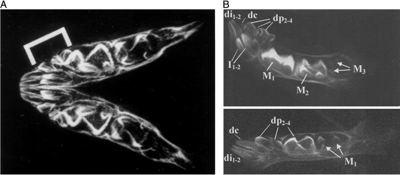
Figure 1.
(A) Radiograph of a mandible of a near-term, captive P. verreauxi from the Natural History Museum (London), specimen no. BMNH 67.1365. The bracket indicates the position of the entire set of deciduous teeth, which are confined to the anterior-most portion of the mandible. M1 and M2 crowns are clearly visible as is the developing crypt for the M3s. (B Upper) Radiograph of a mandible of a newborn (0 days) P. verreauxi coquereli male (Duke University Primate Center specimen DPC 6620M). At birth, the M1 is nearly fully formed, M2 is one-quarter to one-half formed, and the mesial cusps of M3 are visible in the crypt. The deciduous incisors (di1–2) and premolars (dp2–4) are illustrated as are the developing permanent incisors in the region of the symphysis. (Lower) Radiograph of a mandible of a three-day old male Eulemur mongoz (DPC 6537M) showing the deciduous incisors and canine (together comprising the tooth comb) and the anterior deciduous premolars only just erupting. The dp4 is only just one-half crown complete, and only the cusps of the permanent M1 have begun to mineralize.

