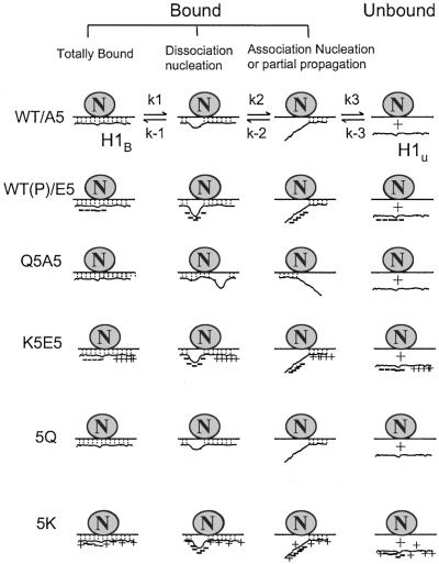
Figure 4.
The nucleation-propagation model of H1-DNA interactions. H1 is proposed to be constantly exchanging, with bound and unbound fractions in equilibrium (30, 31). For all forms of H1, dissociation is nucleated from the region with the lowest positive charge and then propagated to other parts of the molecule. Similarly, association is nucleated from the region with the highest positive charges. In WT cells with fully dephosphorylated H1 or in A5 cells, nucleation of dissociation is relatively low, whereas nucleation of reassociation is high. When WT H1 is phosphorylated or the phosphorylation sites are mutated to glutamic acid (E5), the phosphorylation region is negatively charged and becomes the nucleation center for dissociation. In the Q5A5 series, reducing positive charges creates new nucleation centers in other parts of H1, again shifting the equilibrium toward dissociation. In the K5E5 series, although a nucleation site is produced by the five Es at the phosphorylation sites, we hypothesize that the newly introduced positive charge patches could produce a nucleation center that increases the association rate or that the newly introduced positive charges could produce a kinetic barrier to propagation. In 5Q and 5K cells, the introduced charges are spread out throughout the whole molecule. Because no new nucleation center was created, they behave like A5 or E5 cells, respectively.

