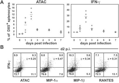
Figure 2.
Correlated secretion of IFN-γ with ATAC, MIP-1α, MIP-1β, and RANTES by NK cells in the innate phase of L. monocytogenes infection. (A) Time course of ATAC and IFN-γ secretion in splenic DX5+ cells. BALB/c mice were infected with L. monocytogenes (five mice per group), and the spleens were removed at various time points. Splenocytes were incubated with brefeldin A for 5 h without restimulation and analyzed for the expression of ATAC and IFN-γ by flow cytometry. Shown is the percentage of DX5+ cells secreting ATAC or IFN-γ in the course of listeriosis. (B) On day 2 (d2) p.i. splenocytes from individual Listeria-infected BALB/c mice were stained for DX5 and counterstained for IFN-γ versus ATAC, MIP-1α, MIP-1β, or RANTES. The percentage of cells in each quadrant and the correlation coefficient φ for each staining pair are indicated. Analysis of one representative animal out of eight is shown.

