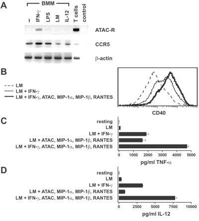
Figure 4.
Functional effects of the type 1 chemokines and IFN-γ on macrophages. (A) BMMs were exposed to IFN-γ, lipopolysaccharide (LPS), or IL-12 or were infected with live L. monocytogenes as described in Materials and Methods. After 24 h of culture, total RNA was analyzed by RT-PCR for the ATAC receptor, CCR5, and β-actin. RNA from purified (99%) CD8+ T cells (T cells) was used as a positive control, and samples without mRNA template were used as a negative control (control). (B) BMMs were infected with Listeria and were either left untreated (LM, dashed line) or treated additionally with IFN-γ alone (thin line) or a combination of IFN-γ and ATAC, MIP-1α, MIP-1β, and RANTES (thick line). After 24 h, BMMs were collected and analyzed for CD40 cell surface expression by using flow cytometry. One representative experiment out of five is shown. (C and D) BMMs were either left untreated (resting) or infected with L. monocytogenes for 1 h. Infected BMMs were either cultured in medium or stimulated with cytokines as indicated. After 24 h, supernatants were harvested and tested for the presence of TNF-α (C) and IL-12 (D). The values represent means of duplicates (TNF-α and IL-12) ± SD. One representative out of four experiments is shown.

