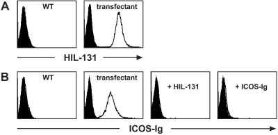
Figure 1.
Characterization of the ICOS-L mAb HIL-131. (A) Binding specificity of HIL-131. Wild-type and ICOS-L-transfected L cells were stained with PE-labeled mAb HIL-131 (open profiles) or an isotype control (filled profiles). (B) Blocking capacity of HIL-131. Wild-type and ICOS-L-transfected L cells were stained with a PE-coupled human ICOS-rabbit-Ig fusion protein (2 μg/ml, open profiles) or a control fusion protein (CD3δ-rabbit-Ig-PE, filled profiles). Staining was blocked by preincubation of the cells with either mAb HIL-131 (10 μg/ml) or an excess of unlabeled human ICOS-rabbit-Ig (100 μg/ml). WT, wild type.

